



Immunohistochemistry (Formalin/PFA-fixed paraffin-embedded sections)
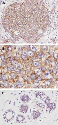
AKT1 (phospho S473) polyclonal antibody (Cat # PAB10323) was used at a 1 : 100 dilution to detect AKT1 by immunohistochemistry of formalin-fixed and paraffin embedded in (A) human breast tumor tissue, (B) a higher magnification and (C) normal human breast tissue of the breast tumor.
The staining is much stronger than the weak basal level of phosphorylation in normal breast.
AKT1 (phospho S473) polyclonal antibody (Cat # PAB10323) was used with no pretreatment of tissue.
Signal was developed using Dako's Techmate streptavidin-biotin reagents.
Immunofluorescence
Immunofluorescence microscopy.
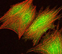
Confocal image using AKT1 (phospho S473) polyclonal antibody (Cat # PAB10323) at a 1 : 40 dilution to detect phosphorylated AKT1 (green) in rat cardiomyocytes infected with adenovirus expressing with wild-type AKT1 in conjunction with a texas-red conjugated phalloidin (red) to label filamentous actin.
Western Blot (Cell lysate)
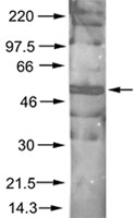
Immunoblotting of AKT1 (phospho S473) polyclonal antibody (Cat # PAB10323) was used at a 1:200 dilution to detect phosphorylated AKT1 by Western blot. A nuclear extract from cells infected with adenovirus expressing nuclear-targeted AKT1 kinase was used.
- Specifications
Product Description
Rabbit polyclonal antibody raised against synthetic phosphopeptide of AKT1.
Immunogen
Synthetic phosphopeptide (conjugated with KLH) corresponding to residues surrounding S473 of human AKT1.
Host
Rabbit
Reactivity
Chicken, Human, Mouse, Rat
Specificity
This antibody is specific to phosphorylated human AKT. Minimal reactivity occurs against non-phosphorylated AKT.
Form
Liquid
Quality Control Testing
Antibody Reactive Against Synthetic Peptide.
Recommend Usage
ELISA (1:15000-1:60000)
Western Blot (1:200-1:1000)
Immunohistochemistry (1:100-1:500)
The optimal working dilution should be determined by the end user.Storage Buffer
In 20 mM KH2PO4, 150 mM NaCl, pH 7.2 (0.01% sodium azide)
Storage Instruction
Store at 4°C. For long term storage store at -20°C.
Aliquot to avoid repeated freezing and thawing. - Applications
Immunohistochemistry (Formalin/PFA-fixed paraffin-embedded sections)
AKT1 (phospho S473) polyclonal antibody (Cat # PAB10323) was used at a 1 : 100 dilution to detect AKT1 by immunohistochemistry of formalin-fixed and paraffin embedded in (A) human breast tumor tissue, (B) a higher magnification and (C) normal human breast tissue of the breast tumor.
The staining is much stronger than the weak basal level of phosphorylation in normal breast.
AKT1 (phospho S473) polyclonal antibody (Cat # PAB10323) was used with no pretreatment of tissue.
Signal was developed using Dako's Techmate streptavidin-biotin reagents.Immunofluorescence
Immunofluorescence microscopy.
Confocal image using AKT1 (phospho S473) polyclonal antibody (Cat # PAB10323) at a 1 : 40 dilution to detect phosphorylated AKT1 (green) in rat cardiomyocytes infected with adenovirus expressing with wild-type AKT1 in conjunction with a texas-red conjugated phalloidin (red) to label filamentous actin.Enzyme-linked Immunoabsorbent Assay
Western Blot (Cell lysate)
Immunoblotting of AKT1 (phospho S473) polyclonal antibody (Cat # PAB10323) was used at a 1:200 dilution to detect phosphorylated AKT1 by Western blot. A nuclear extract from cells infected with adenovirus expressing nuclear-targeted AKT1 kinase was used. - Gene Info — AKT1
Entrez GeneID
207Protein Accession#
P31749;AAH00479Gene Name
AKT1
Gene Alias
AKT, MGC99656, PKB, PKB-ALPHA, PRKBA, RAC, RAC-ALPHA
Gene Description
v-akt murine thymoma viral oncogene homolog 1
Gene Ontology
HyperlinkGene Summary
The serine-threonine protein kinase encoded by the AKT1 gene is catalytically inactive in serum-starved primary and immortalized fibroblasts. AKT1 and the related AKT2 are activated by platelet-derived growth factor. The activation is rapid and specific, and it is abrogated by mutations in the pleckstrin homology domain of AKT1. It was shown that the activation occurs through phosphatidylinositol 3-kinase. In the developing nervous system AKT is a critical mediator of growth factor-induced neuronal survival. Survival factors can suppress apoptosis in a transcription-independent manner by activating the serine/threonine kinase AKT1, which then phosphorylates and inactivates components of the apoptotic machinery. Multiple alternatively spliced transcript variants have been found for this gene. [provided by RefSeq
Other Designations
RAC-alpha serine/threonine-protein kinase|murine thymoma viral (v-akt) oncogene homolog-1|protein kinase B|rac protein kinase alpha
- Interactomes
- Pathways
- Diseases
- Publication Reference
- Cacao powder supplementation attenuates oxidative stress, cholinergic impairment, and apoptosis in D-galactose-induced aging rat brain.
Hyoeun Yoo, Hyun-Sook Kim.
Scientific Reports 2021 Sep; 11(1):17914.
Application:WB, Rat, Rat Brain.
- PKB/Akt: a key mediator of cell proliferation, survival and insulin responses?
Lawlor MA, Alessi DR.
Journal of Cell Science 2001 Aug; 114(Pt 16):2903.
Application:IHC, WB-Tr, Human, Cancers, Mammalian cells.
- Cacao powder supplementation attenuates oxidative stress, cholinergic impairment, and apoptosis in D-galactose-induced aging rat brain.
- Literature Request
Pab, MaxPab, & DNAxPab