Dynasore [304448-55-3]

Référence T1848-5mg
Conditionnement : 5mg
Marque : TargetMol
Contactez votre distributeur local :
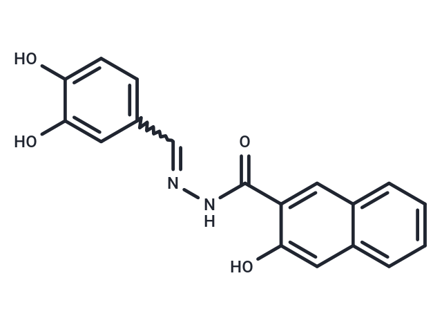
Dynasore
(Synonyms: Dynamin Inhibitor I) Copy Product Info
Dynasore (Dynamin Inhibitor I) is a highly selective, reversible small-molecule dynamin inhibitor with an IC₅₀ of 15 μM. Dynasore also inhibits the mitochondrial dynamin Drp1 without affecting other small GTPases. By inhibiting the GTPase activity of dynamin, Dynasore blocks its function in membrane fission, thereby inhibiting clathrin-mediated endocytosis. Dynasore can be used in research on endocytosis, membrane transport, viral entry, and receptor signaling regulation.
Cas No. 304448-55-3
For research use only—not for human use. No sales to individuals. Use as intended only.
Select Batch
Purity:99.22%
Color:White to Yellow
COA LCMS HNMR HPLC
Product Introduction
Bioactivity
Chemical Properties
Storage & Solubility Information
| Description | Dynasore (Dynamin Inhibitor I) is a highly selective, reversible small-molecule dynamin inhibitor with an IC₅₀ of 15 μM. Dynasore also inhibits the mitochondrial dynamin Drp1 without affecting other small GTPases. By inhibiting the GTPase activity of dynamin, Dynasore blocks its function in membrane fission, thereby inhibiting clathrin-mediated endocytosis. Dynasore can be used in research on endocytosis, membrane transport, viral entry, and receptor signaling regulation. |
| Targets & IC50 | Dynamin1/2:15 μM, moderate potenc:~15 μM (vitro) |
| In vitro | Methods: Mouse bone marrow-derived macrophages were pretreated with 5, 10, 25, or 50 μM Dynasore for 1 hour, then stimulated with 100 ng/mL LPS for 24 hours. Supernatants and cell lysates were collected. Pro-inflammatory cytokine expression levels were detected by real-time quantitative PCR. Results: Dynasore dose-dependently inhibited LPS-induced mRNA and protein levels of proinflammatory cytokines (TNF-α, IL-6, IL-12). [1] Methods: HCLE cells (Fluo-4 loaded) were pretreated with 40 μM Dynasore or 1 μM YM-58483 for 30 min, followed by induction of oxidative stress with 1 mM tBHP. Cytoplasmic calcium changes were monitored in real-time via fluorescence intensity over 2 hours. Results: Dynasore completely blocked tBHP-induced increases in cytoplasmic calcium. [2] Methods: Horizontal brainstem slices containing the solitary tract and solitary tract nucleus were prepared from adult rats. Whole-cell patch-clamp recordings were performed on NTS neurons at a clamp voltage of -60 mV. 100 μM Dynasore dissolved in artificial cerebrospinal fluid was continuously administered via a perfusion system. Postsynaptic currents were recorded at different time points (pre-administration, during administration, post-washout). Results: Dynasore rapidly increased the frequency of spontaneous excitatory postsynaptic currents (EPSCs). Concurrently, ST-evoked EPSCs progressively failed until completely blocked, with no reversal during the recovery period.[3] |
| In vivo | Methods: An acute lung injury (ALI) model was established in C57BL/6J mice via intratracheal LPS (10 mg/kg) administration. Dynasore (10, 30, 50 mg/kg) was administered as a single intraperitoneal injection 3 hours prior to LPS challenge. Mice were euthanized 24 hours after LPS stimulation. Results: Dynasore pretreatment (particularly at 30 and 50 mg/kg) significantly reduced lung tissue histopathology scores and decreased inflammatory mediators. [1] |
| Synonyms | Dynamin Inhibitor I |
| Kinase Assay | RIP1 kinase assay: Phosphorylation of RIP1 requires its kinase activity. Expression constructs of FLAGtagged wild-type (WT) or a kinase-inactive point mutant of RIP1 (K45M) are are transfected into 293T cells and RIP1 kinase assay is performed as described in the Methods in the presence of [γ-32P]ATP for 30 min at 30℃. Samples are subjected to SDS-PAGE and RIP1 band is visualized by autoradiography. Relative intensities of radioactive bands are quantified and are shown (ratio) in this and all other autoradiographs. In parallel to kinase reactions, a sample of beads is subjected to western blot analysis using anti-RIP1 antibody to ensure equal protein amounts in kinase reactions. |
| Cell Research | Mouse ventricular myocytes are isolated from male adult C6/Black mouse. Cardiomyocytes subjects to 2 hours of drug treatment followed by oxidative stress (30 μM Water2 for 35 min). For ATP supplement experiments, the cells are treated with 3 mM ATP for 30 min before exposure to Water2. Cardiomyocyte survival and viability are analyzed by trypan blue exclusion (TBE) assay[3]. |
| Molecular Weight | 322.31 |
| Formula | C18H14N2O4 |
| Cas No. | 304448-55-3 |
| Smiles | Oc1ccc(C=NNC(=O)c2cc3ccccc3cc2O)cc1O |
| Relative Density. | 1.36g/cm3 |
| Storage | Powder: -20°C for 3 years | In solvent: -80°C for 1 year Shipping with blue ice/Shipping at ambient temperature. | ||||||||||||||||||||||||||||||||||||||||
| Solubility Information | Ethanol: 1.6 mg/mL (4.96 mM), Sonication is recommended. DMSO: 120 mg/mL (372.31 mM), Sonication is recommended.  | ||||||||||||||||||||||||||||||||||||||||
| In Vivo Formulation | 10% DMSO+40% PEG300+5% Tween 80+45% Saline: 4 mg/mL (12.41 mM), Sonication is recommended. Please add the solvents sequentially, clarifying the solution as much as possible before adding the next one. Dissolve by heating and/or sonication if necessary. Working solution is recommended to be prepared and used immediately. The formulation provided above is for reference purposes only. In vivo formulations may vary and should be modified based on specific experimental conditions. | ||||||||||||||||||||||||||||||||||||||||
Solution Preparation Table | |||||||||||||||||||||||||||||||||||||||||
Ethanol/DMSO
DMSO
Note : The dilution table applies only to solid products. For liquid products, please calculate the stock solution based on the stated concentration and/or density. | |||||||||||||||||||||||||||||||||||||||||