

Histone H1, Phosphorylated (HRP)

Référence MBS6480516-5x0,1mL
Conditionnement : 5x0.1mL
Marque : MyBiosource
Contactez votre distributeur local :

Téléphone : +1 850 650 7790
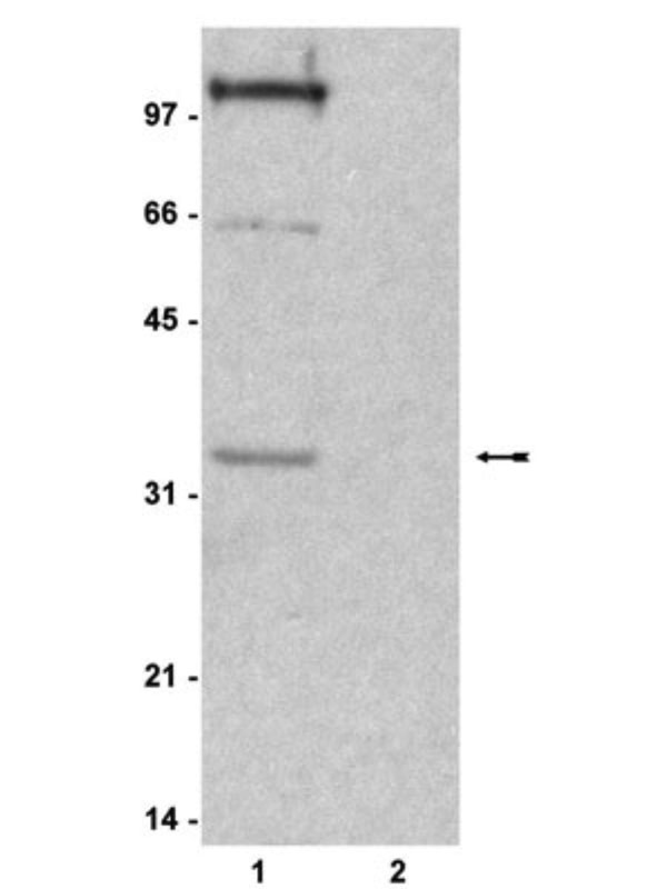
WB: Colcemid treated (lane 1) and untreated (lane 2) HeLa cell lysate probed with MBS628895 (2ug/ml).
Histone H1 (HIST1H1C) Antibody – Rabbit Polyclonal
For Research Use Only. Not for use in diagnostic procedures. Catalog #: MBS6480516MBS6480516 is an antibody targeting HIST1H1C. Also known as Histone H1, Phosphorylated (HRP). Reactive in Human. Host: Rabbit. More details
Product Overview
Online Datasheet in PDFProduct Name
Histone H1 (HIST1H1C), Polyclonal Antibody
Full Product Name
Histone H1, Phosphorylated (HRP)
Product Gene Name
anti-HIST1H1C antibody
Clonality
Polyclonal
Isotype
IgG
Host
Rabbit
Species Reactivity
Human
Form/Format
Supplied as a liquid in PBS, pH 7.2. No preservative added. Labeled with Horseradish Peroxidase (HRP).
Purity/Purification
Purified by Protein A Affinity Chromatography.
Specificity
Recognizes phosphorylated histone H1 (32kD). Additional proteins (Mr 66 and 100kD) were detected. Species Crossreactivity: human
Preparation and Storage
Store product at 4 degree C if to be used immediately within two weeks. For long-term storage, aliquot to avoid repeated freezing and thawing and store at -20 degree C. Aliquots are stable at -20 degree C for 12 months after receipt. Dilute required amount only prior to immediate use. Further dilutions can be made in assay buffer. Note: Sodium azide is a potent inhibitor of peroxidase and should not be added to HRP conjugates.
Applications Tested/Suitable for anti-HIST1H1C antibody
Immunocytochemistry (ICC), Western Blot (WB)
Application Notes for anti-HIST1H1C antibody
ICC: 5-10ug/ml showed positive immunostaining for Histone H1 in mitotic HeLa cells fixed with 95% ethanol, 5% glacial acetic acid.
Applications are based on unconjugated antibody.
WB: 0.5-2ug/ml detected phosphorylated Histone H1 from acid extracts of colcemid treated HeLa cells. Does not detect Histone H1 in untreated HeLa cells. Acid extracts from colcemid treated and untreated HeLa cell were resolved by electrophoresis, transferred to nitrocellulose and probed with. Proteins were visualized using an IgG (HRP) goat anti-rabbit. A chemiluminescence detection system was used.
Applications are based on unconjugated antibody.
WB: 0.5-2ug/ml detected phosphorylated Histone H1 from acid extracts of colcemid treated HeLa cells. Does not detect Histone H1 in untreated HeLa cells. Acid extracts from colcemid treated and untreated HeLa cell were resolved by electrophoresis, transferred to nitrocellulose and probed with. Proteins were visualized using an IgG (HRP) goat anti-rabbit. A chemiluminescence detection system was used.
Product References and Citations for anti-HIST1H1C antibody
1. Dedon, P.C., et al., Anal. Biol. Chem. 197: 83-90, 1991. 2. Pennisi, E., Science 275: 155-157, 1997. 3. Morita, E., et al., J. Virol. 15: 7555-7563, 2001. 4. Bhattacharjee, R., et al., Mol. Cell. Biol. 21: 5417-5425, 2001. 5. Lu, M.J., et al., Chromosoma 103: 111-121, 1994.
Other Notes
Small volumes of anti-HIST1H1C antibody vial(s) may occasionally become entrapped in the seal of the product vial during shipment and storage. If necessary, briefly centrifuge the vial on a tabletop centrifuge to dislodge any liquid in the container`s cap. Certain products may require to ship with dry ice and additional dry ice fee may apply.
Additional Details
NCBI/Uniprot data below describe general gene information for HIST1H1C. It may not necessarily be applicable to this product.
UniProt Primary Accession #
UniProt Secondary Accession #
UniProt Related Accession #
GenBank Nucleotide #
NCBI Official Full Name
histone H1.2
NCBI Official Synonym Full Names
histone cluster 1 H1 family member c
NCBI Official Symbol
HIST1H1C
NCBI Official Synonym Symbols
H1C; H1.2; H1F2; H1s-1
UniProt Protein Name
Histone H1.2
UniProt Synonym Protein Names
Histone H1c; Histone H1d; Histone H1s-1
Protein Family
Histone H1-like protein
UniProt Gene Name
HIST1H1C
UniProt Synonym Gene Names
H1F2
NCBI Protein Information
histone H1.2
NCBI Summary for HIST1H1C
Histones are basic nuclear proteins responsible for nucleosome structure of the chromosomal fiber in eukaryotes. Two molecules of each of the four core histones (H2A, H2B, H3, and H4) form an octamer, around which approximately 146 bp of DNA is wrapped in repeating units, called nucleosomes. The linker histone, H1, interacts with linker DNA between nucleosomes and functions in the compaction of chromatin into higher order structures. This gene is intronless and encodes a replication-dependent histone that is a member of the histone H1 family. Transcripts from this gene lack polyA tails but instead contain a palindromic termination element. This gene is found in the large histone gene cluster on chromosome 6. [provided by RefSeq, Aug 2015]
Show more UniProt Comments for HIST1H1C
Histone H1 protein binds to linker DNA between nucleosomes forming the macromolecular structure known as the chromatin fiber. Histones H1 are necessary for the condensation of nucleosome chains into higher-order structured fibers. Acts also as a regulator of individual gene transcription through chromatin remodeling, nucleosome spacing and DNA methylation ().
Show more Precautions
All of MyBioSource's Products are for scientific laboratory research purposes and are not for diagnostic, therapeutics, prophylactic or in vivo use. Through your purchase, you expressly represent and warrant to MyBioSource that you will properly test and use any Products purchased from MyBioSource in accordance with industry standards. MyBioSource and its authorized distributors reserve the right to refuse to process any order where we reasonably believe that the intended use will fall outside of our acceptable guidelines.
Disclaimer
While every efforts were made to ensure the accuracy of the information provided in this datasheet, MyBioSource will not be liable for any omissions or errors contained herein. MyBioSource reserves the right to make changes to this datasheet at any time without prior notice.
It is the responsibility of the customer to report product performance issues to MyBioSource within 30 days of receipt of the product. Please visit our Terms & Conditions page for more information.
It is the responsibility of the customer to report product performance issues to MyBioSource within 30 days of receipt of the product. Please visit our Terms & Conditions page for more information.
Product Note
Select online data sheet information is drawn from bioinformatics databases, occasionally resulting in ambiguous or non-relevant product information. It is the responsibility of the customer to review, verify, and evaluate the information to make sure it matches their requirements before purchasing the kit. Our ELISA Kit assays are dynamic research tools and sometimes they may be updated and improved. If the format of this assay is important to you then please request the current manual or contact our technical support team with a presales inquiry before placing an order. We will confirm the current details of the assay. We cannot guarantee the sample manual posted online is the most current manual, it is intended to serve as an example only. Please refer to the instructions For Use provided with the assay kit for precise details.
Product PubMed Publications
Products associated with anti-HIST1H1C antibody
| Reference Product | PubMed Publications |
|---|---|
| CDK1 antibody | >1 publications with HIST1H1C and CDK1 |
Pathways associated with anti-HIST1H1C antibody
| Products by Pathway | Pathway Diagram |
|---|---|
| Activation Of DNA Fragmentation Factor Pathway antibodies | Activation Of DNA Fragmentation Factor Pathway Diagram |
| Apoptosis Pathway antibodies | Apoptosis Pathway Diagram |
| Apoptosis Induced DNA Fragmentation Pathway antibodies | Apoptosis Induced DNA Fragmentation Pathway Diagram |
| Apoptotic Execution Phase Pathway antibodies | Apoptotic Execution Phase Pathway Diagram |
| Cellular Senescence Pathway antibodies | Cellular Senescence Pathway Diagram |
| Cellular Responses To Stress Pathway antibodies | Cellular Responses To Stress Pathway Diagram |
| DNA Damage/Telomere Stress Induced Senescence Pathway antibodies | DNA Damage/Telomere Stress Induced Senescence Pathway Diagram |
| Formation Of Senescence-Associated Heterochromatin Foci (SAHF) Pathway antibodies | Formation Of Senescence-Associated Heterochromatin Foci (SAHF) Pathway Diagram |
| Programmed Cell Death Pathway antibodies | Programmed Cell Death Pathway Diagram |
Diseases associated with anti-HIST1H1C antibody
| Disease Name | Pubmed Publications |
|---|---|
| Heart Failure Antibodies | >1 publications with HIST1H1C and Heart Failure |
| Leukemia Antibodies | >1 publications with HIST1H1C and Leukemia |
| Heart Diseases Antibodies | >1 publications with HIST1H1C and Heart Diseases |
| Pancreatic Neoplasms Antibodies | >1 publications with HIST1H1C and Pancreatic Neoplasms |
| Kidney Diseases Antibodies | >1 publications with HIST1H1C and Kidney Diseases |
Organs/Tissues associated with anti-HIST1H1C antibody
| Organ/Tissue Name | Pubmed Publications |
|---|---|
| Blood Antibodies | >2 publications with HIST1H1C and Blood |
| Kidney Antibodies | >1 publications with HIST1H1C and Kidney |
| Heart Antibodies | >1 publications with HIST1H1C and Heart |
| Pancreas Antibodies | >1 publications with HIST1H1C and Pancreas |
| Bone Antibodies | >1 publications with HIST1H1C and Bone |