
HLA-B Antibody - FITC Conjugated

Référence OASA03538
Conditionnement : 0.1mg
Marque : Aviva Systems Biology
Contactez votre distributeur local :

Téléphone : +1 850 650 7790
HLA-B Antibody - FITC Conjugated (OASA03538)

| Datasheets/Manuals | Printable datasheet for HLA-B Antibody - FITC Conjugated (OASA03538) |
|---|
| Predicted Species Reactivity | Mouse | ||||||||||
|---|---|---|---|---|---|---|---|---|---|---|---|
| Product Format | Liquid. Phosphate buffered saline | ||||||||||
| Clonality | Monoclonal | ||||||||||
| Clone | 2G5 | ||||||||||
| Isotype | IgG2b | ||||||||||
| Host | Mouse | ||||||||||
| Conjugation | FITC | ||||||||||
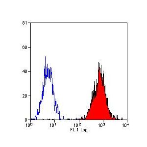
| Application | FC | ||||||||||
| Additional Information | Fusion Partners: Spleen cells from immunised C1D mice were fused with cells of the X63 myeloma cell line. | ||||||||||
| :: | Preparation: Purified IgG prepared by affinity chromatography on Protein G from tissue culture supernatant Preservative Stabilisers: 0.09% - Sodium Azide 1% - Bovine Serum Albumin | ||||||||||
| :: | Approx Protein Conc: IgG concentration 0.1 mg/ml Buffer Solutions: Phosphate buffered saline pH7.4 | ||||||||||
| Reconstitution and Storage | -20°C | ||||||||||
| Immunogen | Purified H-2Kb and H-2Db MHC-I molecules. | ||||||||||
| Purification | Protein A Purified | ||||||||||
| Predicted Homology Based on Immunogen Sequence | Mouse | ||||||||||
| Concentration | 0.1 mg/ml | ||||||||||
| Specificity | MHC CLASS I | ||||||||||
| Application Info | Flow Cytometry: Neat - 1/5 following applications. This information is derived from testing within our laboratories, peer-reviewed publications or personal communications from the originators. Please refer to references indicated for further information.
Where this antibody has not been tested for use in a particular technique this does not necessarily exclude its use in such procedures. Suggested working dilutions are given as a guide only. It is recommended that the user titrates the antibody for use in their own system using appropriate negative\positive controls. | ||||||||||
| Protocol Information | Citation: 1: Huang YC, Hung SW, Jan TR, Liao KW, Cheng CH, Wang YS, Chu RM. CD5-lowexpression lymphocytes in canine peripheral blood show characteristics of naturalkiller cells. J Leukoc Biol. 2008 Dec;84(6):1501-10. Epub 2008 Aug 15. PubMedPMID: 18708592. Species: Beagles Experiment Name: 1. Flow cytometry analysis2. Detection of intracellular IFN- Experiment Background: NK cell markers and receptors have been discovered in many mammalian species, such as humans, mice, rats, pigs, and cows. However, there is still a lack of information concerning NK cell markers or receptors in canines. Yi-Chun et al. have discovered that canine CD5-low density (CD5lo) cells in PBL are closely associated with NK cell characteristics. CD5lo cells comprised 14.9 ± 6.68% of the total PBL. A high proportion of the CD5lo cell population expressed CD3 (96.6%), CD8α (77.7%), CD8β (53%), α/β TCR (83%), and CD11/18 (80%), but the expression of / TCR (6.5%), CD4 (10.6%), and CD21 (2.4%) was low. CD5lo cells were larger than CD5-high density (CD5hi) cells. Light and electron microscopy revealed numerous large cytoplasmic granules in CD5lo cells, especially after IL-2 stimulation, which was in contrast to CD5hi, in which intracytoplasmic granules were not frequently seen. After IL-2 stimulation, CD5lo cells had significantly stronger NK cytotoxicity than CD5hi cells. CD5lo cells had much higher mRNA levels for NKG2D, CD16, CD94, CD160, perforin, and granzyme than CD5hi. Following IL-2 stimulation, CD5lo cells had significantly higher mRNA levels of NKp30, NKp44, CD16, and CD94 than CD5hi cells. In addition, IL-2-stimulated, CD5lo-depleted PBL showed a loss of NK cytotoxicity. CD5lo cells also showed significantly lower antigen-specific cytotoxic T cell activity as compared with CD5hi cells. Taken together, the CD5lo subset in canine PBL is closely related to canine NK cells, and CD5lo can be used as a phenotypic marker for an IL-2-dependent canine NK cell enrichment. Experimental Steps: 1. Flow cytometry analysis: Cells were stained as described previously [25]. Briefly, for direct immunofluorescence analysis, cells were incubated with FITC-conjugated isotype control or FITC-conjugated rat anti-canine CD5 mAb (Oxford, UK). For indirect immunofluorescence analysis, cells were incubated with isotype control or specific mAb raised against CD3, CD4, CD8α, CD8β, CD21, α/β TCR, / TCR, CD14, MHC class I, MHC class II, or CD11/18. Cells were washed and further stained with PE-conjugated goat anti-mouse IgG (Becton Dickinson, San Jose, CA, USA) for 30 min. Finally, all cells were washed and suspended in FACS buffer [2% FBS and 0.02% sodium azide in PBS (pH 7.4)]. The surface immunofluorescence of 104 viable cells was measured with a FACSCaliburTM flow cytometer (Becton Dickinson). Data were analyzed using CellQuest software (Becton Dickinson).2. Detection of intracellular IFN-: Canine PBMCs were analyzed for intracellular IFN- by a flow cytometer. Briefly, PBMCs were incubated in recombinant canine (rc)IL-12 (5 ng/ml; R&D System, Minneapolis, MN, USA) and were collected after 72 h. The cells were then processed for cell surface staining using FITC-conjugated anticanine CD5 mAb. Intracellular staining was performed using anti-IFN--PE (CC302; Oxford, UK) with a BD Cytofix/Cytoperm Plus kit and GolgiStop (Becton Dickinson), according to the manufacturer’s instructions. Cells (10,000)/sample were analyzed in a FACSCaliburTM flow cytometer. Other Reagents Used: RPMI-1640, penicillin, streptomycin, 2-ME, glutamine Number Of Protocols: 2 |

| Reference | 1. Claesson, M.H. et al. (1994) Antibodies directed against monomorphic and evolutionary conserved self epitopes may be generated in 'knock-out' mice. Development of monoclonal antibodies directed against monomorphic MHC class I determinants. Scand. J. Immunol. 40: 257-264. 2. Vitadello, M. et al. (201) Myofiber stress-response in myositis: parallel investigations on patients and experimental animal models of muscle regeneration and systemic inflammation. |
|---|---|
| Gene Symbol | HLA-B |
| Gene Full Name | MHC class I antigen |
| Protein Name | MHC class I antigen |
| Description of Target | MOUSE ANTI MOUSE MHC CLASS I:FITC |
| Uniprot ID | H7CHW7 |
| Protein Size (# AA) | 236 |