
MAPK1 Detection Antibody

Référence OAOA21966
Conditionnement : 50µg
Marque : Aviva Systems Biology
Contactez votre distributeur local :

Téléphone : +1 850 650 7790
MAPK1 Detection Antibody : Biotin (OAOA21966)
| Datasheets/Manuals | Printable datasheet for MAPK1 Detection Antibody : Biotin (OAOA21966) |
|---|
| Predicted Species Reactivity | Human|Mouse|Rat |
|---|---|
| Product Format | Stored in PBS (pH 7.4) with 0.05% sodium azide, 10mg/ml BSA, 50% glycerol |
| Clonality | Monoclonal |
| Clone | OTI4D10 |
| Isotype | IgG1 |
| Host | Mouse |
| Conjugation | Biotin |
| Application | ELISA|MlpxA |
| Additional Information | Matched ELISA Pair: OAOA21505 |
| Reconstitution and Storage | -20°C |
| Immunogen | Full length human recombinant protein of human MAPK1 (NM_138957) produced in HEK293T cell. |
| Concentration | 0.5 mg/ml |
| Dilution | 1:250 - 1:1000 |
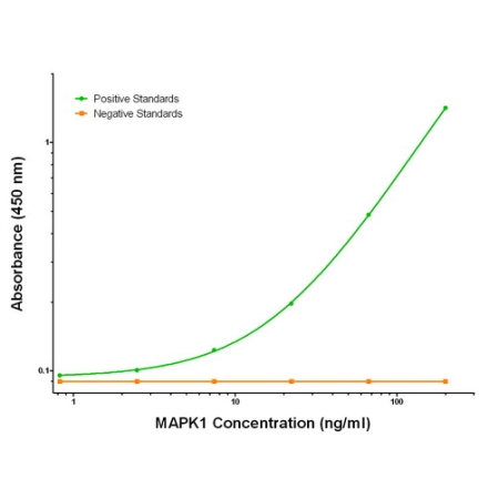
| Description | MAPK1 biotinylated mouse monoclonal detection antibody, validated for ELISA and Luminex assays |
| Gene Symbol | MAPK1 |
|---|---|
| Gene Full Name | mitogen-activated protein kinase 1 |
| Alias Symbols | ERK, ERK-2, ERK2, ERT1, extracellular signal-regulated kinase 2, MAP kinase 1, MAP kinase 2, MAP kinase isoform p42, MAPK 2, MAPK2, mitogen-activated protein kinase 1, mitogen-activated protein kinase 2, NS13, p38, p40, p41, p41mapk, p42-MAPK, P42MAPK, PRKM1, PRKM2, protein tyrosine kinase ERK2. |
| NCBI Gene Id | 5594 |
| Protein Name | Homo sapiens mitogen-activated protein kinase 1 (MAPK1), transcript variant 2, mRNA|Mitogen-activated protein kinase 1 |
| Description of Target | Serine/threonine kinase which acts as an essential component of the MAP kinase signal transduction pathway. MAPK1/ERK2 and MAPK3/ERK1 are the 2 MAPKs which play an important role in the MAPK/ERK cascade. They participate also in a signaling cascade initiated by activated KIT and KITLG/SCF. Depending on the cellular context, the MAPK/ERK cascade mediates diverse biological functions such as cell growth, adhesion, survival and differentiation through the regulation of transcription, translation, cytoskeletal rearrangements. The MAPK/ERK cascade plays also a role in initiation and regulation of meiosis, mitosis, and postmitotic functions in differentiated cells by phosphorylating a number of transcription factors. About 160 substrates have already been discovered for ERKs. Many of these substrates are localized in the nucleus, and seem to participate in the regulation of transcription upon stimulation. However, other substrates are found in the cytosol as well as in other cellular organelles, and those are responsible for processes such as translation, mitosis and apoptosis. Moreover, the MAPK/ERK cascade is also involved in the regulation of the endosomal dynamics, including lysosome processing and endosome cycling through the perinuclear recycling compartment (PNRC); as well as in the fragmentation of the Golgi apparatus during mitosis. The substrates include transcription factors (such as ATF2, BCL6, ELK1, ERF, FOS, HSF4 or SPZ1), cytoskeletal elements (such as CANX, CTTN, GJA1, MAP2, MAPT, PXN, SORBS3 or STMN1), regulators of apoptosis (such as BAD, BTG2, CASP9, DAPK1, IER3, MCL1 or PPARG), regulators of translation (such as EIF4EBP1) and a variety of other signaling-related molecules (like ARHGEF2, DCC, FRS2 or GRB10). Protein kinases (such as RAF1, RPS6KA1/RSK1, RPS6KA3/RSK2, RPS6KA2/RSK3, RPS6KA6/RSK4, SYK, MKNK1/MNK1, MKNK2/MNK2, RPS6KA5/MSK1, RPS6KA4/MSK2, MAPKAPK3 or MAPKAPK5) and phosphatases (such as DUSP1, DUSP4, DUSP6 or DUSP16) are other substrates which enable the propagation the MAPK/ERK signal to additional cytosolic and nuclear targets, thereby extending the specificity of the cascade. Mediates phosphorylation of TPR in respons to EGF stimulation. May play a role in the spindle assembly checkpoint. Phosphorylates PML and promotes its interaction with PIN1, leading to PML degradation. Phosphorylates CDK2AP2 (By similarity). |
| Uniprot ID | P28482 |
| Protein Accession # | NP_620407 |
| Nucleotide Accession # | https://www.ncbi.nlm.nih.gov/nuccore/NM_138957 |