
Mouse CD5/Ly-1 Antibody : FITC

Référence OASB00287
Conditionnement : 0.1mg
Marque : Aviva Systems Biology
Contactez votre distributeur local :

Téléphone : +1 850 650 7790
Mouse CD5/Ly-1 Antibody : FITC (OASB00287)
| Datasheets/Manuals | Printable datasheet for Mouse CD5/Ly-1 Antibody : FITC (OASB00287) |
|---|
| Tested Species Reactivity | Mouse |
|---|---|
| Predicted Species Reactivity | Mouse |
| Clonality | Monoclonal |
| Clone | 4H8E5 |
| Isotype | IgG2a |
| Host | Rat |
| Conjugation | FITC |
| Application | FC |
| Additional Information | Description: CD5, also known as Lyt-1, is a monomeric type I transmembrane glycoprotein expressed on thymocytes, T lymphocytes, and a subset of B lymphocytes, but not on natural killer (NK) cells. It has been identified as the major ligand of the B-cell antigen CD72. The frequency of CD5+ B cells exhibits strain-dependent variation, and the phenotypic, anatomical, functional, developmental, and pathological characteristics of the CD5+ B cells suggest that they may represent a distinct lineage, known as B-1 cells. Binding of CD5 on the T cell surface can augment alloantigen- or mitogen-induced lymphocyte proliferation and induces increased cytosolic free calcium, IL-2 secretion, and IL-2R expression. It has been proposed that CD5 negatively regulates signal transduction mediated by the T-cell and B-cell receptors. |
| Reconstitution and Storage | Store at 2-8C |
| Concentration | 0.1 mg/mL |
| Specificity | CD5 |
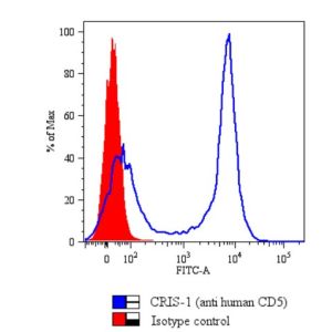
| Characterization | To ensure lot- to- lot consistency, each batch of monoclonal antibody is tested to conform withcharacteristics of a standard by flow cytometry. Representative data are included in this data sheet. |
| Warning | Reagents contain sodium azide. Sodium azide is very toxic if ingested or inhaled. Avoid contact with skin,eyes, or clothing. Wear eye or face protection when handling. If skin or eye contact occurs, wash withcopious amounts of water. If ingested or inhaled, contact a physician immediately. Sodium azide yieldstoxic hydrazoic acid under acidic conditions. Dilute azide- containing compounds in running water beforediscarding to avoid accumulation of potentially explosive deposits in lead or copper plumbing. |
| Dilution | Flow Cytometry: Fluorescein conjugate <= 1 ug/106 cells Biotin conjugate <= 1 ug/106 cells R-phycoerythrin conjugate Allophycocyanin conjugate Spectral Red conjugate <= 0.2 ug/106 cells <= 0.2 ug/106 cells <= 0.2 ug/106 cells |
| Application Info | Identification and enumeration of CD5+ cells by flow cytometry 15 |
| Other Applications Data | Since applications vary, you should determine the optimum working dilution for the product that is appropriate for yourspecific need. |
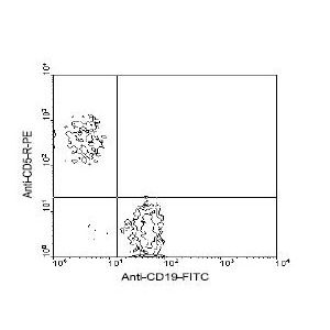
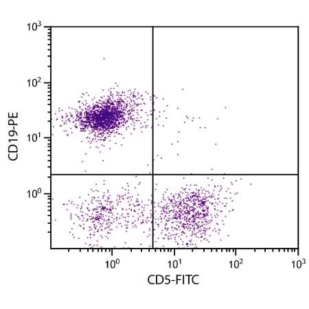
| Other Applications Image 1 Data | Amount Used: ≤ 0.2 Ug/106 cells Cells from BALB/c spleen were double-stained with rat anti-mouse CD5-R-PE and rat anti-mouse CD19-FITC , following which small lymphocytes were gated and analyzed using a FACScan flow cytometer (BDIS, San Jose, CA) |
| Storage | - The purified (UNLB) antibody is supplied as 0.5 mg of purified immunoglobulin in 1.0 mL of 100 mMborate buffered saline, pH 8.2. No preservatives or amine- containing buffer salts added. Store at 2- 8 C - The fluorescein (FITC) conjugate is supplied as 0.5 mg or 0.1 mg in 1.0 mL of PBS/NaN3. Store at 2- 8 C - The biotin (BIOT) conjugate is supplied as 0.5 mg in 1.0 mL of PBS/NaN3. Store at 2- 8 C - The R- phycoerythrin (R- PE) conjugate is supplied as 0.1 mg in 1.0 mL or 0.2 mg in 2.0 mL ofPBS/NaN3 and a stabilizing agent. Store at 2- 8 C. Do not freeze! - The allophycocyanin (APC) conjugate is supplied as 0.1 mg in 1.0 mL of PBS/NaN3 and a stabilizingagent. Store at 2- 8 C. Do not freeze! - The Spectral Red (SPRD) conjugate is supplied as 0.1 mg in 1.0 mL of PBS/NaN3 and a stabilizingagent. Store at 2- 8 C. Do not freeze! - Protect conjugated forms from light. Each reagent is stable for the period shown on the bottle label ifstored as directed. |
| Reference | 1. Ledbetter, J. A. and L. A. Herzenberg. 1979. Immunol. Rev. 47:63 2. Seaman, W. E., D. Wofsy, J. S. Greenspan, and J. A. Ledbetter. 1983. J. Immunol. 130:1713 3. Ledbetter, J. A., R. V. Rouse, H. S. Micklem, and L. A. Herzenberg. 1980. J. Exp. Med. 152:280 4. Van de Veld, H., I. von Hoegen, W. Luo, J. R. Parnes, and K. Thielemans. 1991. Nature 351:662 5. Luo, W., H. Van de Velde, I. von Hoegen, J. R. Parnes, and K. Thielemans. 1992. J. Immunol. 148:1630 6. Kantor, A. B. and L. A. Herzenberg. 1993. Ann. Rev. Immunol. 11:501 7. Holland, N. 1982. Immunol. Rev. 68:43 8. Stanton, T., T. L. Stevens, J. A. Ledbetter, and D. Wofsy. 1986. J. Immunol. 136:1734 9. Verwilghen J, R. Vandesande, P. Vandenberghe, J.L. Ceuppens . 1990. Cell Immunol. 131:109 10. Logdberg, L. and E. M. Shevach. 1996. Eur. J. Immunol. 15:1007 11. June, C. H., P. R. Rabinovitch, and J. A. Ledbetter. 1987. J. Immunol. 138:2782 12. Hollander, N., E. Pillemer, and I. L. Weissman. 1981. Proc. Natl. Acad. Sci. U. S. A. 78:1148 13. Tarakhovsky, A., S.B. Kanner, J. Hombach, J.A. ledbetter, W. Muller, N. Killeen, and K. Rajewsky. 1995. Science 269:535 14. Bikah, G., J. Carey, J.R. Cialella, A. Tarakhovsky, and S, Bobdada. 1996. Science 274:1906 15. Saunderson,S.C., P.C. Schuberth, A.C. Dunn, L. Miller, B.D. Hock, P.A. MacKay, N. Koch, R.W. Jack, and A.D. McLellan 2008. J. Immunol. 180:8146. |
|---|---|
| Gene Symbol | CD5 |
| Gene Full Name | T-cell surface glycoprotein CD5 |
| Alias Symbols | Ly, Ly-, Ly-1, Ly-A, Lyt-, Ly-12, Lyt-1 |
| NCBI Gene Id | 12507 |
| Protein Name | T-cell surface glycoprotein CD5 |
| Uniprot ID | P13379 |
| Protein Accession # | NP_031676.3 |
| Nucleotide Accession # | NM_007650.3 |