

p53 Leeporter HeLa Cell Line

Référence MBS668869-1
Conditionnement : 1Vial
Marque : MyBiosource
Contactez votre distributeur local :

Téléphone : +1 850 650 7790
p53 Leeporter HeLa Cell Line
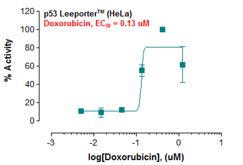
Fig-1: Induction of p53 activity by doxorubicin in p53 Leeporter HeLa cells.
p53 Cell Line
For Research Use Only. Not for use in diagnostic procedures. Catalog #: MBS668869The p53 Leeporter cell line is a stably transfected HeLa cell line which expresses Renilla luciferase reporter gene under the transcriptional control of the p53 response element. p53 is a tumor suppressor that plays a crucial role in apoptosis and anticancer… More details
Product Overview
Online Datasheet in PDFProduct Name
p53, Cell Line
Full Product Name
p53 Leeporter HeLa Cell Line
Product Gene Name
p53 cell line
Form/Format
Each vial contains 2 ~ 3 x 10^6 cells in 1 ml of 90% FBS + 10% DMSO.
Preparation and Storage
Immediately upon receipt, store in liquid nitrogen.
Dry Ice Shipment: Extra charge fee may add to your shipping cost as dry ice is required to ship this product.
Shipping Note: Product is available for shipment in the United States, Canada and European countries. Please inquire for shipment to other countries.
Dry Ice Shipment: Extra charge fee may add to your shipping cost as dry ice is required to ship this product.
Shipping Note: Product is available for shipment in the United States, Canada and European countries. Please inquire for shipment to other countries.
Molecular Weight
24,401 Da
OMIM
X02469 mRNA
Dry Ice Shipment
Extra charge fee may add to your shipping cost as dry ice is required to ship this product.
Product Categories/Family for p53 cell line
Cell Lines; Reporter Cell Lines
Applications Tested/Suitable for p53 cell line
Functional Assay
Application Notes for p53 cell line
Monitor the p53 induction activity. Screen for activators or inhibitors of the p53 signaling pathway. Culture conditions: Cells should be grown at 37 degree C with 5% CO2 using DMEM medium supplemented with 10% FBS and 1% Pen/Strep, plus 3 ug/ml of Puromycin. It is recommended to quickly thaw the frozen cells upon receipt or from liquid nitrogen in a 37 degree C water-bath, transfer to a tube containing 10 ml of growth medium without Puromycin, spin down cells, resuspend cells in pre-warmed growth medium without Puromycin, transfer resuspended cells to T25 flask and culture in 37 degree C-CO2 incubator. Leave the T25 flask in the incubator for 2~4 days without disturbing or changing the medium until cells completely recover viability and become adherent. Once cells are over 90% adherent, remove growth medium and passage the cells through trypsinization and centrifugation. At first passage, switch to growth medium containing Puromycin. Cells should be split before they reach complete confluence. To passage the cells, detach cells from culture vessel with Trypsin/EDTA, add complete growth medium and transfer to a tube, spin down cells, resuspend cells and seed appropriate aliquots of cells suspension into new culture vessels. Subcultivation ration = 1: 10 to 1: 20 weekly. Functional validation: A. Response of p53 Leeporter- HeLa cells to doxorubicin. 1. Harvest p53 Leeporter- HeLa cells and seed cells into a white solid-bottom 96-well microplate in 100 ul of growth medium at 5 x 10^4 cells/well. 2. Incubate cells at 37 degree C in a CO2 incubator for overnight. 3. The next day, stimulate cells with various concentrations of doxorubicin. 4. Incubate at 37 degree C in a CO2 incubator for 6-16 hours. 5. Add 30-50 ul of luciferase assay reagent per well. 6. Incubate at room temperature for 1-5 minutes and measure luminescence using a microplate luminometer.
Other Notes
Small volumes of p53 cell line vial(s) may occasionally become entrapped in the seal of the product vial during shipment and storage. If necessary, briefly centrifuge the vial on a tabletop centrifuge to dislodge any liquid in the container`s cap. Certain products may require to ship with dry ice and additional dry ice fee may apply.
Searchable Terms for p53 purchase
MBS668869 is a cell line targeting p53. Also known as p53 Leeporter HeLa Cell Line.
Additional Details
NCBI/Uniprot data below describe general gene information for p53. It may not necessarily be applicable to this product.
NCBI Official Full Name
P53
NCBI Official Synonym Full Names
tumor protein p53
NCBI Official Symbol
TP53
NCBI Official Synonym Symbols
P53; BCC7; LFS1; TRP53
UniProt Protein Name
Cellular tumor antigen p53
UniProt Synonym Protein Names
Antigen NY-CO-13; Phosphoprotein p53; Tumor suppressor p53
Protein Family
p53 and DNA damage-regulated protein
UniProt Gene Name
TP53
UniProt Synonym Gene Names
P53
NCBI Protein Information
cellular tumor antigen p53
NCBI Summary for p53
This gene encodes a tumor suppressor protein containing transcriptional activation, DNA binding, and oligomerization domains. The encoded protein responds to diverse cellular stresses to regulate expression of target genes, thereby inducing cell cycle arrest, apoptosis, senescence, DNA repair, or changes in metabolism. Mutations in this gene are associated with a variety of human cancers, including hereditary cancers such as Li-Fraumeni syndrome. Alternative splicing of this gene and the use of alternate promoters result in multiple transcript variants and isoforms. Additional isoforms have also been shown to result from the use of alternate translation initiation codons from identical transcript variants (PMIDs: 12032546, 20937277). [provided by RefSeq, Dec 2016]
Show more UniProt Comments for p53
p53: a transcription factor and major tumor suppressor that plays a major role in regulating cellular responses to DNA damage and other genomic aberrations. Activation of p53 can lead to either cell cycle arrest and DNA repair or apoptosis. More than 50 percent of human tumors contain a mutation or deletion of the TP53 gene. p53 is modified post-translationally at multiple sites. DNA damage induces phosphorylation of p53 at S15, S20 and S37, reducing its interaction with the oncoprotein MDM2. MDM2 inhibits p53 accumulation by targeting it for ubiquitination and proteasomal degradation. Phosphorylated by many kinases including Chk2 and Chk1 at S20, enhancing its tetramerization, stability and activity. The phosphorylation by CAK at S392 is increased in human tumors and has been reported to influence the growth suppressor function, DNA binding and transcriptional activation of p53. Phosphorylation of p53 at S46 regulates the ability of p53 to induce apoptosis. The acetylation of p53 appears to play a positive role in the accumulation of p53 during the stress response. Following DNA damage, p53 becomes acetylated at K382, enhancing its binding to DNA. Deacetylation of p53 can occur through interaction with SIRT1, a deacetylase that may be involved in cellular aging and the DNA damage response. p53 regulates the transcription of a set of genes encoding endosomal proteins that regulate endosomal functions. These include STEAP3 and CHMP4C, which enhance exosome production, and CAV1 and CHMP4C, which produce a more rapid endosomal clearance of the EGFR from the plasma membrane. DNA damage regulates a p53-mediated secretory pathway, increasing the secretion of some proteins such as Hsp90, SERPINE1, SERPINB5, NKEF-A, and CyPA, and inhibiting the secretion of others including CTSL and IGFBP-2. Two alternatively spliced human isoforms have been reported. Isoform 2 is expressed in quiescent lymphocytes. Seems to be non-functional. May be produced at very low levels due to a premature stop codon in the mRNA, leading to nonsense-mediated mRNA decay.
Protein type: Activator; DNA-binding; Motility/polarity/chemotaxis; Nuclear receptor co-regulator; Transcription factor; Tumor suppressor
Chromosomal Location of Human Ortholog: 17p13.1
Cellular Component: cytoplasm; cytosol; mitochondrion; nuclear body; nuclear chromatin; nuclear matrix; nucleolus; nucleoplasm; nucleus; PML body; protein complex; replication fork; transcription factor TFIID complex
Molecular Function: ATP binding; chaperone binding; chromatin binding; copper ion binding; damaged DNA binding; DNA binding; double-stranded DNA binding; enzyme binding; histone acetyltransferase binding; identical protein binding; p53 binding; protease binding; protein binding; protein heterodimerization activity; protein kinase binding; protein N-terminus binding; protein phosphatase 2A binding; protein phosphatase binding; protein self-association; receptor tyrosine kinase binding; sequence-specific DNA binding; transcription factor activity; transcription factor binding; ubiquitin protein ligase binding; zinc ion binding
Biological Process: base-excision repair; cell aging; cell cycle arrest; cell differentiation; cell proliferation; cellular response to glucose starvation; chromatin assembly; circadian behavior; determination of adult life span; DNA damage response, signal transduction by p53 class mediator; DNA damage response, signal transduction by p53 class mediator resulting in cell cycle arrest; DNA damage response, signal transduction by p53 class mediator resulting in induction of apoptosis; DNA damage response, signal transduction by p53 class mediator resulting in transcription of p21 class mediator; DNA strand renaturation; entrainment of circadian clock by photoperiod; ER overload response; G1 DNA damage checkpoint; multicellular organismal development; negative regulation of apoptosis; negative regulation of cell growth; negative regulation of cell proliferation; negative regulation of fibroblast proliferation; negative regulation of helicase activity; negative regulation of telomerase activity; negative regulation of transcription from RNA polymerase II promoter; negative regulation of transcription, DNA-dependent; nucleotide-excision repair; positive regulation of apoptosis; positive regulation of histone deacetylation; positive regulation of neuron apoptosis; positive regulation of peptidyl-tyrosine phosphorylation; positive regulation of protein export from nucleus; positive regulation of protein oligomerization; positive regulation of transcription from RNA polymerase II promoter; positive regulation of transcription, DNA-dependent; proteasomal ubiquitin-dependent protein catabolic process; protein complex assembly; protein localization; protein sumoylation; protein tetramerization; Ras protein signal transduction; regulation of apoptosis; regulation of mitochondrial membrane permeability; regulation of transcription, DNA-dependent; response to antibiotic; response to DNA damage stimulus; response to gamma radiation; response to X-ray; viral reproduction
Disease: Adrenocortical Carcinoma, Hereditary; Basal Cell Carcinoma, Susceptibility To, 7; Breast Cancer; Colorectal Cancer; Glioma Susceptibility 1; Hepatocellular Carcinoma; Li-fraumeni Syndrome 1; Nasopharyngeal Carcinoma; Osteogenic Sarcoma; Pancreatic Cancer; Papilloma Of Choroid Plexus
Show more Protein type: Activator; DNA-binding; Motility/polarity/chemotaxis; Nuclear receptor co-regulator; Transcription factor; Tumor suppressor
Chromosomal Location of Human Ortholog: 17p13.1
Cellular Component: cytoplasm; cytosol; mitochondrion; nuclear body; nuclear chromatin; nuclear matrix; nucleolus; nucleoplasm; nucleus; PML body; protein complex; replication fork; transcription factor TFIID complex
Molecular Function: ATP binding; chaperone binding; chromatin binding; copper ion binding; damaged DNA binding; DNA binding; double-stranded DNA binding; enzyme binding; histone acetyltransferase binding; identical protein binding; p53 binding; protease binding; protein binding; protein heterodimerization activity; protein kinase binding; protein N-terminus binding; protein phosphatase 2A binding; protein phosphatase binding; protein self-association; receptor tyrosine kinase binding; sequence-specific DNA binding; transcription factor activity; transcription factor binding; ubiquitin protein ligase binding; zinc ion binding
Biological Process: base-excision repair; cell aging; cell cycle arrest; cell differentiation; cell proliferation; cellular response to glucose starvation; chromatin assembly; circadian behavior; determination of adult life span; DNA damage response, signal transduction by p53 class mediator; DNA damage response, signal transduction by p53 class mediator resulting in cell cycle arrest; DNA damage response, signal transduction by p53 class mediator resulting in induction of apoptosis; DNA damage response, signal transduction by p53 class mediator resulting in transcription of p21 class mediator; DNA strand renaturation; entrainment of circadian clock by photoperiod; ER overload response; G1 DNA damage checkpoint; multicellular organismal development; negative regulation of apoptosis; negative regulation of cell growth; negative regulation of cell proliferation; negative regulation of fibroblast proliferation; negative regulation of helicase activity; negative regulation of telomerase activity; negative regulation of transcription from RNA polymerase II promoter; negative regulation of transcription, DNA-dependent; nucleotide-excision repair; positive regulation of apoptosis; positive regulation of histone deacetylation; positive regulation of neuron apoptosis; positive regulation of peptidyl-tyrosine phosphorylation; positive regulation of protein export from nucleus; positive regulation of protein oligomerization; positive regulation of transcription from RNA polymerase II promoter; positive regulation of transcription, DNA-dependent; proteasomal ubiquitin-dependent protein catabolic process; protein complex assembly; protein localization; protein sumoylation; protein tetramerization; Ras protein signal transduction; regulation of apoptosis; regulation of mitochondrial membrane permeability; regulation of transcription, DNA-dependent; response to antibiotic; response to DNA damage stimulus; response to gamma radiation; response to X-ray; viral reproduction
Disease: Adrenocortical Carcinoma, Hereditary; Basal Cell Carcinoma, Susceptibility To, 7; Breast Cancer; Colorectal Cancer; Glioma Susceptibility 1; Hepatocellular Carcinoma; Li-fraumeni Syndrome 1; Nasopharyngeal Carcinoma; Osteogenic Sarcoma; Pancreatic Cancer; Papilloma Of Choroid Plexus
Precautions
All of MyBioSource's Products are for scientific laboratory research purposes and are not for diagnostic, therapeutics, prophylactic or in vivo use. Through your purchase, you expressly represent and warrant to MyBioSource that you will properly test and use any Products purchased from MyBioSource in accordance with industry standards. MyBioSource and its authorized distributors reserve the right to refuse to process any order where we reasonably believe that the intended use will fall outside of our acceptable guidelines.
Disclaimer
While every efforts were made to ensure the accuracy of the information provided in this datasheet, MyBioSource will not be liable for any omissions or errors contained herein. MyBioSource reserves the right to make changes to this datasheet at any time without prior notice.
It is the responsibility of the customer to report product performance issues to MyBioSource within 30 days of receipt of the product. Please visit our Terms & Conditions page for more information.
It is the responsibility of the customer to report product performance issues to MyBioSource within 30 days of receipt of the product. Please visit our Terms & Conditions page for more information.
Product Note
Select online data sheet information is drawn from bioinformatics databases, occasionally resulting in ambiguous or non-relevant product information. It is the responsibility of the customer to review, verify, and evaluate the information to make sure it matches their requirements before purchasing the kit. Our ELISA Kit assays are dynamic research tools and sometimes they may be updated and improved. If the format of this assay is important to you then please request the current manual or contact our technical support team with a presales inquiry before placing an order. We will confirm the current details of the assay. We cannot guarantee the sample manual posted online is the most current manual, it is intended to serve as an example only. Please refer to the instructions For Use provided with the assay kit for precise details.
Product PubMed Publications
Pathways associated with p53 cell line
| Products by Pathway | Pathway Diagram |
|---|---|
| AMPK Signaling Pathway antibodies | AMPK Signaling Pathway Diagram |
| Activation Of BH3-only Proteins Pathway antibodies | Activation Of BH3-only Proteins Pathway Diagram |
| Activation Of NOXA And Translocation To Mitochondria Pathway antibodies | Activation Of NOXA And Translocation To Mitochondria Pathway Diagram |
| Activation Of PUMA And Translocation To Mitochondria Pathway antibodies | Activation Of PUMA And Translocation To Mitochondria Pathway Diagram |
| Alzheimers Disease Pathway antibodies | Alzheimers Disease Pathway Diagram |
| Amyotrophic Lateral Sclerosis (ALS) Pathway antibodies | Amyotrophic Lateral Sclerosis (ALS) Pathway Diagram |
| Amyotrophic Lateral Sclerosis (ALS) Pathway antibodies | Amyotrophic Lateral Sclerosis (ALS) Pathway Diagram |
| Apoptosis Pathway antibodies | Apoptosis Pathway Diagram |
| Apoptosis Pathway antibodies | Apoptosis Pathway Diagram |
| Apoptosis Pathway antibodies | Apoptosis Pathway Diagram |
Diseases associated with p53 cell line
| Disease Name | Pubmed Publications |
|---|---|
| Carcinoma Antibodies | >2213 publications with p53 and Carcinoma |
| Urogenital Neoplasms Antibodies | >960 publications with p53 and Urogenital Neoplasms |
| Breast Neoplasms Antibodies | >869 publications with p53 and Breast Neoplasms |
| Colorectal Neoplasms Antibodies | >601 publications with p53 and Colorectal Neoplasms |
| Carcinoma, Squamous Cell Antibodies | >530 publications with p53 and Carcinoma, Squamous Cell |
| Glioma Antibodies | >467 publications with p53 and Glioma |
| Cell Transformation, Neoplastic Antibodies | >327 publications with p53 and Cell Transformation, Neoplastic |
| Carcinoma, Small Cell Antibodies | >319 publications with p53 and Carcinoma, Small Cell |
| Neoplasm Metastasis Antibodies | >289 publications with p53 and Neoplasm Metastasis |
| Sarcoma Antibodies | >278 publications with p53 and Sarcoma |
Organs/Tissues associated with p53 cell line
| Organ/Tissue Name | Pubmed Publications |
|---|---|
| Blood Antibodies | >715 publications with p53 and Blood |
| Lung Antibodies | >581 publications with p53 and Lung |
| Brain Antibodies | >489 publications with p53 and Brain |
| Liver Antibodies | >298 publications with p53 and Liver |
| Skin Antibodies | >260 publications with p53 and Skin |
| Lymph Antibodies | >234 publications with p53 and Lymph |
| Prostate Antibodies | >181 publications with p53 and Prostate |
| Vascular Antibodies | >160 publications with p53 and Vascular |
| Muscle Antibodies | >119 publications with p53 and Muscle |
| Kidney Antibodies | >110 publications with p53 and Kidney |