

Phospho-DAPK1 (Ser734) Antibody

Référence MBS9614140-5x0,2mL
Conditionnement : 5x0.2mL
Marque : MyBiosource
Contactez votre distributeur local :

Téléphone : +1 850 650 7790
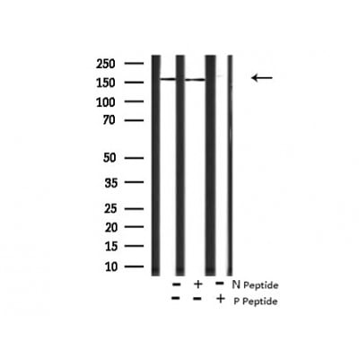
Western blot analysis of DAPK1 (Phospho-Ser734) using HepG2 whole cell lysates.
-/+ means absence or presence of N peptide(non-phospho peptide) and P peptide(phospho peptide).
-/+ means absence or presence of N peptide(non-phospho peptide) and P peptide(phospho peptide).
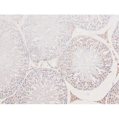
Staining Rat testis tissue sections by IHC-P. The tissue was formaldehyde fixed and a heat mediated antigen retrieval step in citrate buffer was performed. The tissue was then blocked and incubated with the antibody for 1.5 hours at 22 degree C. An HRP conjugated goat anti-rabbit antibody was used as the secondary antibody.
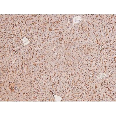
At 1/200 staining Mouse liver tissue sections by IHC-P. The tissue was formaldehyde fixed and a heat mediated antigen retrieval step in citrate buffer was performed. The tissue was then blocked and incubated with the antibody for 1.5 hours at 22 degree C. An HRP conjugated goat anti-rabbit antibody was used as the secondary antibody.
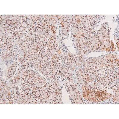
At 1/200 staining Human liver cancer tissue sections by IHC-P. The tissue was formaldehyde fixed and a heat mediated antigen retrieval step in citrate buffer was performed. The tissue was then blocked and incubated with the antibody for 1.5 hours at 22 degree C. An HRP conjugated goat anti-rabbit antibody was used as the secondary antibody.
DAPK1 Antibody – Rabbit Polyclonal
For Research Use Only. Not for use in diagnostic procedures. Catalog #: MBS9614140Calcium/calmodulin-dependent serine/threonine kinase involved in multiple cellular signaling pathways that trigger cell survival, apoptosis, and autophagy. Regulates both type I apoptotic and type II autophagic cell deaths signal, depending on the cellular… More details
Product Overview
Online Datasheet in PDFProduct Name
DAPK1, Polyclonal Antibody
Full Product Name
Phospho-DAPK1 (Ser734) Antibody
Product Gene Name
anti-DAPK1 antibody
Product Synonym Names
DAK1; DAP K1; DAP kinase 1; DAPK 1; DAPK; DAPK1; DAPK1_HUMAN; Death Associated Protein Kinase 1; Death-associated protein kinase 1; DKFZp781I035
Clonality
Polyclonal
Isotype
Rabbit IgG
Host
Rabbit
Species Reactivity
Human, Mouse, Rat
Predicted Reactivity: Pig (91%), Bovine (91%), Horse (89%), Sheep (91%), Rabbit (91%), Dog (91%), Chicken (83%), Xenopus (90%)
Predicted Reactivity: Pig (91%), Bovine (91%), Horse (89%), Sheep (91%), Rabbit (91%), Dog (91%), Chicken (83%), Xenopus (90%)
Form/Format
Liquid. Rabbit IgG in phosphate buffered saline, pH7.4, 150mM NaCl, 0.02% sodium azide and 50% glycerol.
Purity/Purification
The antibody is from purified rabbit serum by affinity purification via sequential chromatography on phospho-peptide and non-phospho-peptide affinity columns.
Concentration
1mg/ml (lot specific)
Specificity
Phospho-DAPK1 (Ser734) Antibody detects endogenous levels of DAPK1 only when phosphorylated at Ser736.
Tissue Specificity: Isoform 2 is expressed in normal intestinal tissue as well as in colorectal carcinomas.
Tissue Specificity: Isoform 2 is expressed in normal intestinal tissue as well as in colorectal carcinomas.
Preparation and Storage
Store at -20 degree C. Stable for 12 months from date of receipt.
Antibody/Peptide Pairs
DAPK1 (Ser734) peptide (MBS9619102) is used for blocking the activity of DAPK1 (Ser734) antibody (MBS9614140)
Molecular Weight
1430
Chromosome Location
Chromosome: 9; NC_000009.11 (90112601..90323566). Location: 9q21.33
OMIM
600831
Supply Chain Verification
Manufactured in a lab with traceable raw materials manufactured on site. Coordinated product portfolio of antibodies, pairs, conjugates, recombinant proteins, and immunoassay materials available, please inquire.
Immunogen
A synthesized peptide derived from human DAPK1 around the phosphorylation site of Ser734.
Conjugation
Unconjugated
Fragment
Fab fragment
Post Translational Modifications
Ubiquitinated by the BCR (KLHL20) E3 ubiquitin ligase complex, leading to its degradation by the proteasome.Removal of the C-terminal tail of isoform 2 (corresponding to amino acids 296-337 of isoform 2) by proteolytic cleavage stimulates maximally its membrane-blebbing function.In response to mitogenic stimulation (PMA or EGF), phosphorylated at Ser-289; phosphorylation suppresses DAPK1 pro-apoptotic function. Autophosphorylation at Ser-308 inhibits its catalytic activity. Phosphorylation at Ser-734 by MAPK1 increases its catalytic activity and promotes cytoplasmic retention of MAPK1. Endoplasmic-stress can cause dephosphorylation at Ser-308.
Subunit Structure
Interacts with KLHL20. Interacts (via death domain) with MAPK1 and MAPK3. Interacts with MAP1B (via N-terminus). Interacts with PRKD1 in an oxidative stress-regulated manner. Interacts with PIN1, PDCD6, BECN1, TSC2 and STX1A. Interacts (via kinase domain) with DAPK3 (via kinase domain). Interacts with GRINB (By similarity). Interacts (via death domain) with UNC5B (via death domain). Interacts with UNC5C (via death domain).
Similarity
The autoinhibitory domain sterically blocks the substrate peptide-binding site by making both hydrophobic and electrostatic contacts with the kinase core.Belongs to the protein kinase superfamily. CAMK Ser/Thr protein kinase family. DAP kinase subfamily.
Subcellular Location
Cytoplasm. Cytoplasm>Cytoskeleton.
Note: Colocalizes with MAP1B in the microtubules and cortical actin fibers.Cytoplasm. Cytoplasm>Cytoskeleton.
Note: Colocalizes with MAP1B in the microtubules and cortical actin fibers.Cytoplasm. Cytoplasm>Cytoskeleton.
ISO Certification
Manufactured in an ISO 9001:2015 Certified Laboratory.
Applications Tested/Suitable for anti-DAPK1 antibody
Western Blot (WB), Immunohistochemistry (IHC), Peptide ELISA (EIA)
Application Notes for anti-DAPK1 antibody
WB: 1:1000-3000
IHC: 1:50-1:200
Peptide ELISA: 1:20,000-1:40,000
IHC: 1:50-1:200
Peptide ELISA: 1:20,000-1:40,000
Immunohistochemistry (IHC) of anti-DAPK1 antibody
Staining Rat testis tissue sections by IHC-P. The tissue was formaldehyde fixed and a heat mediated antigen retrieval step in citrate buffer was performed. The tissue was then blocked and incubated with the antibody for 1.5 hours at 22 degree C. An HRP conjugated goat anti-rabbit antibody was used as the secondary antibody.
Immunohistochemistry (IHC) of anti-DAPK1 antibody
At 1/200 staining Mouse liver tissue sections by IHC-P. The tissue was formaldehyde fixed and a heat mediated antigen retrieval step in citrate buffer was performed. The tissue was then blocked and incubated with the antibody for 1.5 hours at 22 degree C. An HRP conjugated goat anti-rabbit antibody was used as the secondary antibody.
Immunohistochemistry (IHC) of anti-DAPK1 antibody
At 1/200 staining Human liver cancer tissue sections by IHC-P. The tissue was formaldehyde fixed and a heat mediated antigen retrieval step in citrate buffer was performed. The tissue was then blocked and incubated with the antibody for 1.5 hours at 22 degree C. An HRP conjugated goat anti-rabbit antibody was used as the secondary antibody.
Other Notes
Small volumes of anti-DAPK1 antibody vial(s) may occasionally become entrapped in the seal of the product vial during shipment and storage. If necessary, briefly centrifuge the vial on a tabletop centrifuge to dislodge any liquid in the container`s cap. Certain products may require to ship with dry ice and additional dry ice fee may apply.
Searchable Terms for DAPK1 purchase
MBS9614140 is an antibody targeting DAPK1. Also known as Phospho-DAPK1 (Ser734) Antibody. Reactive in Human, Mouse, Rat
Predicted Reactivity: Pig (91%), Bovine (91%), Horse (89%), Sheep (91%), Rabbit (91%), Dog (91%), Chicken (83%), Xenopus (90%). Host: Rabbit.
Predicted Reactivity: Pig (91%), Bovine (91%), Horse (89%), Sheep (91%), Rabbit (91%), Dog (91%), Chicken (83%), Xenopus (90%). Host: Rabbit.
Additional Details
NCBI/Uniprot data below describe general gene information for DAPK1. It may not necessarily be applicable to this product.
NCBI Official Full Name
death-associated protein kinase 1
NCBI Official Synonym Full Names
death-associated protein kinase 1
NCBI Official Symbol
DAPK1
NCBI Official Synonym Symbols
DAPK
UniProt Protein Name
Death-associated protein kinase 1
Protein Family
Death-associated protein kinase
UniProt Gene Name
DAPK1
UniProt Synonym Gene Names
DAPK; DAP kinase 1
UniProt Entry Name
DAPK1_HUMAN
NCBI Protein Information
death-associated protein kinase 1; DAP kinase 1
Precautions
All of MyBioSource's Products are for scientific laboratory research purposes and are not for diagnostic, therapeutics, prophylactic or in vivo use. Through your purchase, you expressly represent and warrant to MyBioSource that you will properly test and use any Products purchased from MyBioSource in accordance with industry standards. MyBioSource and its authorized distributors reserve the right to refuse to process any order where we reasonably believe that the intended use will fall outside of our acceptable guidelines.
Disclaimer
While every efforts were made to ensure the accuracy of the information provided in this datasheet, MyBioSource will not be liable for any omissions or errors contained herein. MyBioSource reserves the right to make changes to this datasheet at any time without prior notice.
It is the responsibility of the customer to report product performance issues to MyBioSource within 30 days of receipt of the product. Please visit our Terms & Conditions page for more information.
It is the responsibility of the customer to report product performance issues to MyBioSource within 30 days of receipt of the product. Please visit our Terms & Conditions page for more information.
Product Note
Select online data sheet information is drawn from bioinformatics databases, occasionally resulting in ambiguous or non-relevant product information. It is the responsibility of the customer to review, verify, and evaluate the information to make sure it matches their requirements before purchasing the kit. Our ELISA Kit assays are dynamic research tools and sometimes they may be updated and improved. If the format of this assay is important to you then please request the current manual or contact our technical support team with a presales inquiry before placing an order. We will confirm the current details of the assay. We cannot guarantee the sample manual posted online is the most current manual, it is intended to serve as an example only. Please refer to the instructions For Use provided with the assay kit for precise details.
Product PubMed Publications
Products associated with anti-DAPK1 antibody
| Reference Product | PubMed Publications |
|---|---|
| CDKN2A antibody | >46 publications with DAPK1 and CDKN2A |
| MAPK3 antibody | >2 publications with DAPK1 and MAPK3 |
| MAPK1 antibody | >2 publications with DAPK1 and MAPK1 |
| HOXB7 antibody | >1 publications with DAPK1 and HOXB7 |
| PDCD6 antibody | >1 publications with DAPK1 and PDCD6 |
Pathways associated with anti-DAPK1 antibody
| Products by Pathway | Pathway Diagram |
|---|---|
| Apoptosis Pathway antibodies | Apoptosis Pathway Diagram |
| Bladder Cancer Pathway antibodies | Bladder Cancer Pathway Diagram |
| Bladder Cancer Pathway antibodies | Bladder Cancer Pathway Diagram |
| IFN-gamma Pathway antibodies | IFN-gamma Pathway Diagram |
| Pathways In Cancer antibodies | Pathways In Cancer Diagram |
| Regulation Of Apoptosis Pathway antibodies | Regulation Of Apoptosis Pathway Diagram |
| Role Of DCC In Regulating Apoptosis Pathway antibodies | Role Of DCC In Regulating Apoptosis Pathway Diagram |
Diseases associated with anti-DAPK1 antibody
| Disease Name | Pubmed Publications |
|---|---|
| Neoplasms Antibodies | >116 publications with DAPK1 and Neoplasms |
| Carcinoma, Squamous Cell Antibodies | >23 publications with DAPK1 and Carcinoma, Squamous Cell |
| Adenocarcinoma Antibodies | >21 publications with DAPK1 and Adenocarcinoma |
| Brain Diseases Antibodies | >19 publications with DAPK1 and Brain Diseases |
| Breast Neoplasms Antibodies | >16 publications with DAPK1 and Breast Neoplasms |
| Lung Neoplasms Antibodies | >13 publications with DAPK1 and Lung Neoplasms |
| Necrosis Antibodies | >12 publications with DAPK1 and Necrosis |
| Carcinoma, Non-Small-Cell Lung Antibodies | >9 publications with DAPK1 and Carcinoma, Non-Small-Cell Lung |
| Leukemia, Myeloid, Acute Antibodies | >9 publications with DAPK1 and Leukemia, Myeloid, Acute |
| Neoplasms, Experimental Antibodies | >9 publications with DAPK1 and Neoplasms, Experimental |