



Recombinant Human IL-1 beta, derived from the transfected human HEK293 cells.
Recombinant Human IL-1 beta Protein, HEK293
Recombinant Human IL-1 beta Protein, HEK293
Contact Us for Bulk Request
Product Description
Specifications
| Size | 10 µg, 50 µg, 100 µg, 250 µg |
| Species | Human |
| Accession Number | P01584 |
| Gene Id | 3553 |
| Gene Symbols | IL1B, IL1F2 |
| Protein Name / Synonyms | Interleukin-1 beta (IL-1 beta) (Catabolin), Interleukin-1 beta, IL-1 beta, Catabolin, Interleukin 1 Beta, IL1F2, Pro-Interleukin-1-Beta, Preinterleukin 1 Beta, IL1-BETA, IL-1 |
| Expressed Region | Ala117-Ser269 |
| Expression System | HEK293 cells |
| Tag | N-terminal histidine tag |
| Purity | >95% |
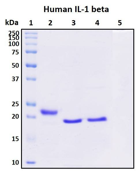
| Purity Determined By | SDS-PAGE under reducing conditions and visualized by Coomassie blue staining |
| Molecular Weight (kDa) | Recombinant protein has a calculated molecular weight of about 17 kDa. Due to the post-translation modifications, it migrates as approximately 25 kDa protein band in SDS-PAGE under DTT, beta-mercaptoethanol reducing conditions. |
| Format | Lyophilized powder |
| Formulation | Lyophilized from a 0.2 µm filtered solution in PBS |
| Concentration (lot specific) | Determined by Pierce BCA protein assay kit |
| Preservative | None |
| Reconstitution | Briefly spin the vial and bring the contents to the bottom prior to opening. It is recommended to reconstitute at 0.5 - 1 mg/mL with sterile deionized water. |
| Endotoxin Level | Not determined |
| Research Area | Angiogenesis, Inflammation |
| Shipping Type | Ambient temperature |
| Storage | -20°C |
Related Products
Recombinant Human IL-1 beta Protein, Biotinylated
As low as $186.00
Introduction
Interleukin-1 Beta (IL-1β) is a pro-inflammatory cytokine predominantly produced by activated macrophages. It plays a pivotal role in mediating immune and inflammatory responses by inducing the expression of various genes involved in inflammation, immunity, and hematopoiesis. IL-1β is synthesized as an inactive precursor and requires cleavage by caspase-1 to become active. Dysregulation of IL-1β production or signaling is associated with various pathological conditions, including autoimmune diseases, chronic inflammatory diseases, and sepsis.
Reference: https://pubmed.ncbi.nlm.nih.gov/8630372/
SDS-PAGE Image
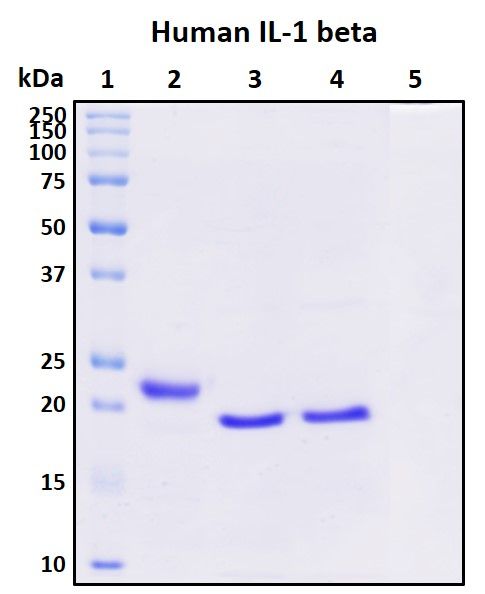
Lane 1: Protein standard ladder (kDa).
Lane 2: Untreated protein under reducing conditions.
Lane 3: Treated protein with deglycosylation enzymes under native conditions.
Lane 4: Treated protein with deglycosylation enzymes under reducing conditions.
Lane 5: Deglycosylation mixture only without target proteins.
Activity
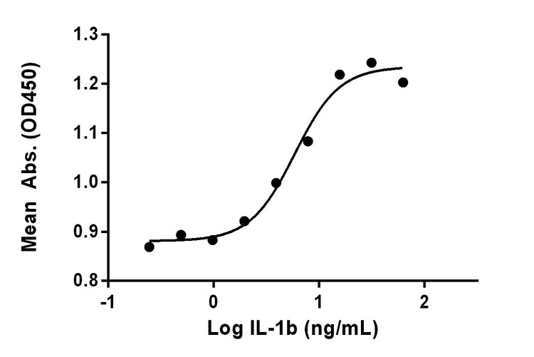
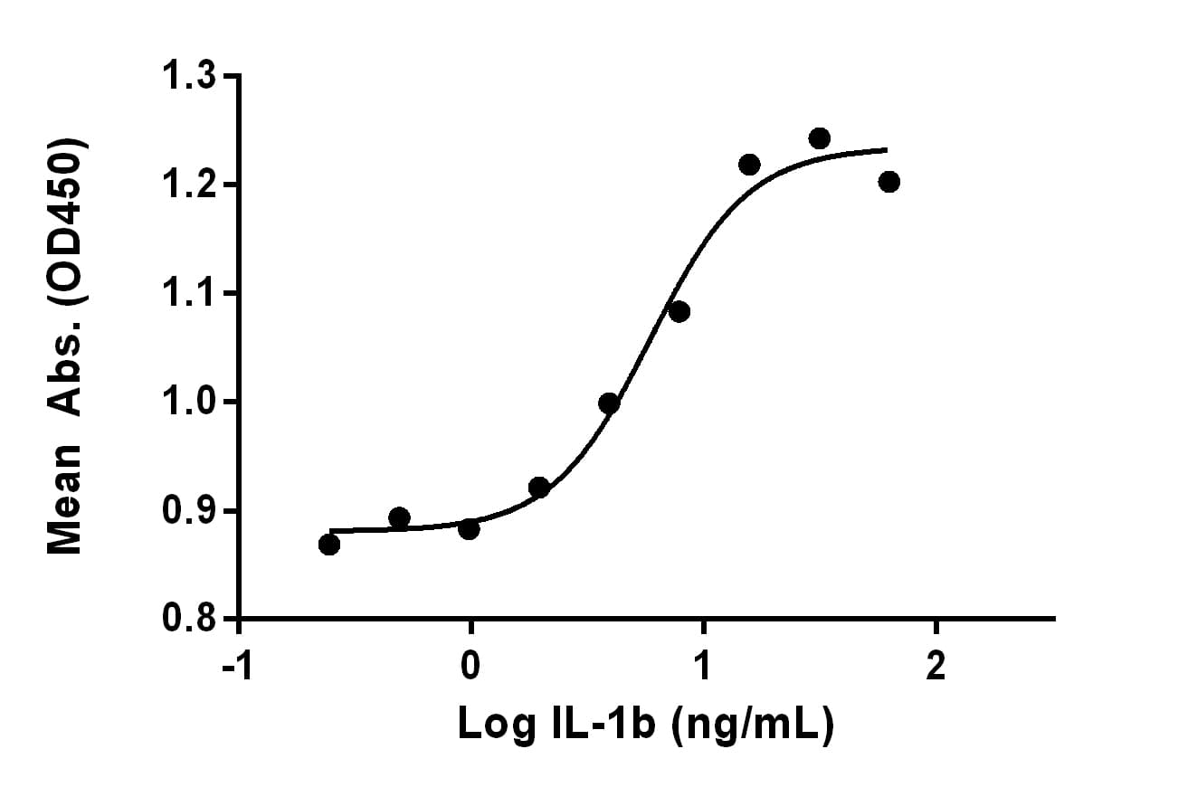
Stimulating cell growth significantly. Measured in a cell proliferation assay using TF-1 human erythroleukemic cells. The EC50 for this effect is 5.8 ng/mL.
Storage/Stability
Upon arrival, the lyophilized protein may be stored for 2 weeks at 4°C. For long term storage, it is recommended to store desiccated below -20°C in a manual defrost freezer. Following reconstitution, the protein may be stored for 2 weeks under sterile conditions at -20°C. For long term storage, it is recommended to make appropriate aliquots and store at -80°C. Avoid repeated freeze-thaw cycles.
Custom Labeling Request
We can custom label this protein with biotin or fluorescent dyes (choose from violet, blue, green or red). Request now.
documents
citations
Transcriptional activation of hepcidin by the microphthalmia/transcription factor E family
- Year:
- 2022
- Journal Name:
- Cell Biochemistry and Function