

IgA Secretory Component (ECM1, Extracellular Matrix Protein 1, Secretory Component p85, URBWD) (AP)

Cat# MBS6124282-5x0,1mL
Size : 5x0.1mL
Brand : MyBiosource
Contact local distributor :

Phone : +1 850 650 7790
IgA Secretory Component (ECM1, Extracellular Matrix Protein 1, Secretory Component p85, URBWD) (AP)
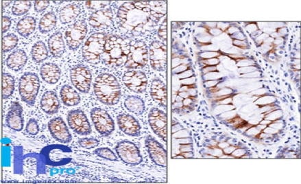
Formalin-fixed, paraffin-embedded normal human small intestine stained with Secretory Component MAb (SPM217). Note cytoplasmic and membrane staining.
Secretory Component (IgA) Secondary Antibody – Mouse Monoclonal
For Research Use Only. Not for use in diagnostic procedures. Catalog #: MBS6124282MBS6124282 is a secondary antibody targeting IgA. Also known as IgA Secretory Component (ECM1, Extracellular Matrix Protein 1, Secretory Component p85, URBWD) (AP). Reactive in Human, Rat. Host: Mouse. More details
Product Overview
Online Datasheet in PDFProduct Name
IgA Secretory Component, Secondary Antibody
Full Product Name
IgA Secretory Component (ECM1, Extracellular Matrix Protein 1, Secretory Component p85, URBWD) (AP)
Product Gene Name
IgA secondary antibody
Product Synonym Names
ECM1; Extracellular Matrix Protein 1; Secretory Component p85; URBWD
Clonality
Monoclonal
Isotype
IgG1,k
Clone Number
SPM217
Host
Mouse
Species Reactivity
Human, Rat
Form/Format
Supplied as a liquid in 10mM PBS. No preservative added. BSA-Free. Labeled with Alkaline Phosphatase (AP).
Purity/Purification
Purified by Protein A/G Affinity Chromatography.
Specificity
Recognizes IgA Secretory Component. Species Crossreactivity: Human and Rat.
Preparation and Storage
May be stored at 4 degree C before opening. DO NOT FREEZE! Stable at 4 degree C as an undiluted liquid. Dilute only prior to immediate use. Stable for 12 months after receipt. For maximum recovery of product, centrifuge the original vial after thawing and prior to removing the cap. Further dilutions can be made in assay buffer. Freezing alkaline phosphatase conjugates will result in a substantial loss of activity.
Molecular Weight
60,674 Da
Chromosome Location
Chromosome: 1; NC_000001.10 (150480487..150486265). Location: 1q21
OMIM
247100
Immunogen
Secretory Component protein isolated from human colostrum. Cellular Localization: Cell surface and cytoplasmic
Conjugate
AP
Positive Control
Stomach, Lung, or breast tumor
Product Categories/Family for IgA secondary antibody
Antibodies; IgA
Applications Tested/Suitable for IgA secondary antibody
ELISA (EIA), Immunofluorescence (IF), Immunohistochemistry (IHC), Immunoprecipitation (IP), Western Blot (WB)
Application Notes for IgA secondary antibody
IP: 1-2ug/500ug protein lysate
IHC: 0.5-1ug/ml for 30 minutes at RT
IF: 1-2ug/ml
WB: 0.5-1ug/ml
Applications are based on unconjugated antibody.
IHC: 0.5-1ug/ml for 30 minutes at RT
IF: 1-2ug/ml
WB: 0.5-1ug/ml
Applications are based on unconjugated antibody.
Product References and Citations for IgA secondary antibody
1. Bartek J et. al. Histochemistry 91:235-244 (1989).
Other Notes
Small volumes of IgA secondary antibody vial(s) may occasionally become entrapped in the seal of the product vial during shipment and storage. If necessary, briefly centrifuge the vial on a tabletop centrifuge to dislodge any liquid in the container`s cap. Certain products may require to ship with dry ice and additional dry ice fee may apply.
Additional Details
NCBI/Uniprot data below describe general gene information for IgA. It may not necessarily be applicable to this product.
NCBI Official Full Name
extracellular matrix protein 1 isoform 3
NCBI Official Synonym Full Names
extracellular matrix protein 1
NCBI Official Symbol
ECM1
NCBI Official Synonym Symbols
URBWD
UniProt Protein Name
Extracellular matrix protein 1
UniProt Synonym Protein Names
Secretory component p85
Protein Family
IgA-specific serine endopeptidase autotransporter
UniProt Gene Name
ECM1
UniProt Entry Name
ECM1_HUMAN
NCBI Protein Information
extracellular matrix protein 1; secretory component p85
NCBI Summary for IgA
This gene encodes a soluble protein that is involved in endochondral bone formation, angiogenesis, and tumor biology. It also interacts with a variety of extracellular and structural proteins, contributing to the maintenance of skin integrity and homeostasis. Mutations in this gene are associated with lipoid proteinosis disorder (also known as hyalinosis cutis et mucosae or Urbach-Wiethe disease) that is characterized by generalized thickening of skin, mucosae and certain viscera. Alternatively spliced transcript variants encoding distinct isoforms have been described for this gene. [provided by RefSeq, Feb 2011]
Show more UniProt Comments for IgA
ECM1: Involved in endochondral bone formation as negative regulator of bone mineralization. Stimulates the proliferation of endothelial cells and promotes angiogenesis. Inhibits MMP9 proteolytic activity. Defects in ECM1 are the cause of lipoid proteinosis (LiP); also known as lipoid proteinosis of Urbach and Wiethe or hyalinosis cutis et mucosae. LiP is a rare autosomal recessive disorder characterized by generalized thickening of skin, mucosae and certain viscera. Classical features include beaded eyelid papules and laryngeal infiltration leading to hoarseness. Histologically, there is widespread deposition of hyaline material and disruption/reduplication of basement membrane. 4 isoforms of the human protein are produced by alternative splicing.
Protein type: Secreted, signal peptide; Secreted
Chromosomal Location of Human Ortholog: 1q21
Cellular Component: extracellular matrix; proteinaceous extracellular matrix; extracellular space
Molecular Function: protein C-terminus binding; protein binding; signal transducer activity; enzyme binding; protease binding; interleukin-2 receptor binding; laminin binding
Biological Process: ossification; positive regulation of I-kappaB kinase/NF-kappaB cascade; negative regulation of peptidase activity; signal transduction; regulation of transcription from RNA polymerase II promoter; negative regulation of bone mineralization; positive regulation of angiogenesis; biomineral formation; negative regulation of cytokine and chemokine mediated signaling pathway; positive regulation of endothelial cell proliferation; angiogenesis; regulation of T-helper 2 type immune response; inflammatory response
Disease: Lipoid Proteinosis Of Urbach And Wiethe
Show more Protein type: Secreted, signal peptide; Secreted
Chromosomal Location of Human Ortholog: 1q21
Cellular Component: extracellular matrix; proteinaceous extracellular matrix; extracellular space
Molecular Function: protein C-terminus binding; protein binding; signal transducer activity; enzyme binding; protease binding; interleukin-2 receptor binding; laminin binding
Biological Process: ossification; positive regulation of I-kappaB kinase/NF-kappaB cascade; negative regulation of peptidase activity; signal transduction; regulation of transcription from RNA polymerase II promoter; negative regulation of bone mineralization; positive regulation of angiogenesis; biomineral formation; negative regulation of cytokine and chemokine mediated signaling pathway; positive regulation of endothelial cell proliferation; angiogenesis; regulation of T-helper 2 type immune response; inflammatory response
Disease: Lipoid Proteinosis Of Urbach And Wiethe
Precautions
All of MyBioSource's Products are for scientific laboratory research purposes and are not for diagnostic, therapeutics, prophylactic or in vivo use. Through your purchase, you expressly represent and warrant to MyBioSource that you will properly test and use any Products purchased from MyBioSource in accordance with industry standards. MyBioSource and its authorized distributors reserve the right to refuse to process any order where we reasonably believe that the intended use will fall outside of our acceptable guidelines.
Disclaimer
While every efforts were made to ensure the accuracy of the information provided in this datasheet, MyBioSource will not be liable for any omissions or errors contained herein. MyBioSource reserves the right to make changes to this datasheet at any time without prior notice.
It is the responsibility of the customer to report product performance issues to MyBioSource within 30 days of receipt of the product. Please visit our Terms & Conditions page for more information.
It is the responsibility of the customer to report product performance issues to MyBioSource within 30 days of receipt of the product. Please visit our Terms & Conditions page for more information.
Product Note
Select online data sheet information is drawn from bioinformatics databases, occasionally resulting in ambiguous or non-relevant product information. It is the responsibility of the customer to review, verify, and evaluate the information to make sure it matches their requirements before purchasing the kit. Our ELISA Kit assays are dynamic research tools and sometimes they may be updated and improved. If the format of this assay is important to you then please request the current manual or contact our technical support team with a presales inquiry before placing an order. We will confirm the current details of the assay. We cannot guarantee the sample manual posted online is the most current manual, it is intended to serve as an example only. Please refer to the instructions For Use provided with the assay kit for precise details.
Product PubMed Publications
Diseases associated with IgA secondary antibody
| Disease Name | Pubmed Publications |
|---|---|
| Lipoid Proteinosis of Urbach and Wiethe Antibodies | >44 publications with IgA and Lipoid Proteinosis of Urbach and Wiethe |
| Neoplasms Antibodies | >27 publications with IgA and Neoplasms |
| Carcinoma Antibodies | >17 publications with IgA and Carcinoma |
| Breast Neoplasms Antibodies | >9 publications with IgA and Breast Neoplasms |
| Colitis, Ulcerative Antibodies | >9 publications with IgA and Colitis, Ulcerative |
| Carcinoma, Squamous Cell Antibodies | >6 publications with IgA and Carcinoma, Squamous Cell |
| Inflammation Antibodies | >6 publications with IgA and Inflammation |
| Adenocarcinoma Antibodies | >5 publications with IgA and Adenocarcinoma |
| Disease Models, Animal Antibodies | >3 publications with IgA and Disease Models, Animal |
| Fibrosis Antibodies | >2 publications with IgA and Fibrosis |
Organs/Tissues associated with IgA secondary antibody
| Organ/Tissue Name | Pubmed Publications |
|---|---|
| Skin Antibodies | >49 publications with IgA and Skin |
| Blood Antibodies | >24 publications with IgA and Blood |
| Vascular Antibodies | >17 publications with IgA and Vascular |
| Bone Antibodies | >10 publications with IgA and Bone |
| Brain Antibodies | >7 publications with IgA and Brain |
| Lymph Node Antibodies | >7 publications with IgA and Lymph Node |
| Muscle Antibodies | >6 publications with IgA and Muscle |
| Embryonic Tissue Antibodies | >5 publications with IgA and Embryonic Tissue |
| Thyroid Antibodies | >5 publications with IgA and Thyroid |
| Connective Tissue Antibodies | >5 publications with IgA and Connective Tissue |