

Fibronectin (Total)

Référence MBS4381665-0,1mg-BSA-AZ
Conditionnement : 0.1mg(WithBSA&Azideat0.2mg/mL)
Marque : MyBiosource
Contactez votre distributeur local :

Téléphone : +1 850 650 7790
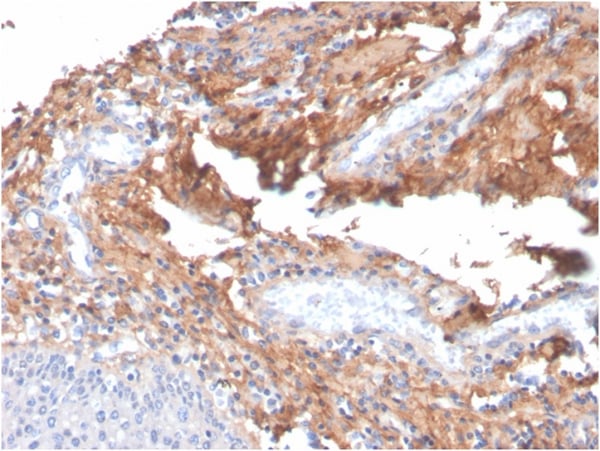
Formalin-fixed, paraffin-embedded human tonsil stained with Fibronectin Mouse Monoclonal Antibody (C6F10).
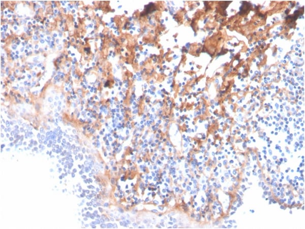
Formalin-fixed, paraffin-embedded human tonsil stained with Fibronectin Mouse Monoclonal Antibody (C6F10).
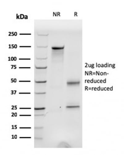
SDS-PAGE Analysis Purified Fibronectin Mouse Monoclonal Antibody (C6F10). Confirmation of Integrity and Purity of Antibody.
Fibronectin (FN1) Antibody – Mouse Monoclonal
For Research Use Only. Not for use in diagnostic procedures. Catalog #: MBS4381665MBS4381665 is an antibody targeting FN1. Also known as Fibronectin (Total). Reactive in Human, Monkey, Porcine, Bovine, Equine, Avian, Amphibian. Host: Mouse. More details
Product Overview
Online Datasheet in PDFProduct Name
Fibronectin (FN1), Monoclonal Antibody
Full Product Name
Fibronectin (Total)
Product Gene Name
anti-FN1 antibody
Product Synonym Names
Cold insoluble globulin (CIG); FINC; FN1; FNZ; GFND; GFND2; LETS; Migration stimulating factor (MSF); Ugl-Y3
Clonality
Monoclonal
Isotype
IgG1, kappa
Clone Number
C6F10
Host
Mouse
Species Reactivity
Human, Monkey, Porcine, Bovine, Equine, Avian, Amphibian
Form/Format
Prepared in 10mM PBS with 0.05% BSA & 0.05% azide.
Purity/Purification
200ug/ml of Ab purified from Bioreactor Concentrate by Protein A/G.
Specificity
Fibronectin is an extracellular matrix glycoprotein present on most cell surfaces, in extracellular fluids and in plasma. A high molecular weight heterodimeric protein, it was originally discovered as a protein missing from the surfaces of virus-transformed cells, and it has been shown to be involved in various functions including cell adhesion, cell motility and wound healing. Alternative splicing and glycosylation give rise to several different forms of Fibronectin, some of which exhibit restricted tissue distribution or association with malignancies. It has been shown that myofibroblast phenotype formation correlates with the occurrence of glycosylated Fibronectin and Fibronectin splice variants in Dupuytrens disease.
Preparation and Storage
Antibody with azide: store at 2 to 8 degree C. Non-hazardous. No MSDS required.
Applications Tested/Suitable for anti-FN1 antibody
Immunofluorescence (IF), Immunohistochemistry (IHC), Western Blot (WB)
Application Notes for anti-FN1 antibody
WB: 2-4µg/ml
IF: 1-3µg/ml
IHC: 1-2µg/ml for 30 minutes at RT
Staining of formalin-fixed tissues requires heating tissue sections in 10mM Tris with 1mM EDTA, pH 9.0, for 45 min at 95 degree C followed by cooling at RT for 20 minutes
Optimal dilution for a specific application should be determined.
IF: 1-3µg/ml
IHC: 1-2µg/ml for 30 minutes at RT
Staining of formalin-fixed tissues requires heating tissue sections in 10mM Tris with 1mM EDTA, pH 9.0, for 45 min at 95 degree C followed by cooling at RT for 20 minutes
Optimal dilution for a specific application should be determined.
Product References and Citations for anti-FN1 antibody
Ugarova et. al. Biochemistry, 1996; 35:10913-10921. | Shi, Z., et al. 2010. The neuroprotective effect of Batch-2, an aqueous extract from cats claw (Uncaria tomentosa) on 6-OHDA-induced SH-SY5Y cell damage. Prog. Biochem. Biophys. 37: 769-778.
Other Notes
Small volumes of anti-FN1 antibody vial(s) may occasionally become entrapped in the seal of the product vial during shipment and storage. If necessary, briefly centrifuge the vial on a tabletop centrifuge to dislodge any liquid in the container`s cap. Certain products may require to ship with dry ice and additional dry ice fee may apply.
Additional Details
NCBI/Uniprot data below describe general gene information for FN1. It may not necessarily be applicable to this product.
NCBI Official Full Name
fibronectin isoform 3 preproprotein
NCBI Official Synonym Full Names
fibronectin 1
NCBI Official Symbol
FN1
NCBI Official Synonym Symbols
FN; CIG; FNZ; MSF; ED-B; FINC; GFND; LETS; GFND2; SMDCF
UniProt Protein Name
Fibronectin
UniProt Synonym Protein Names
Cold-insoluble globulin
Protein Family
Fibronectin
UniProt Gene Name
FN1
UniProt Synonym Gene Names
FN; FN; CIG
UniProt Entry Name
FINC_HUMAN
NCBI Protein Information
fibronectin
NCBI Summary for FN1
This gene encodes fibronectin, a glycoprotein present in a soluble dimeric form in plasma, and in a dimeric or multimeric form at the cell surface and in extracellular matrix. The encoded preproprotein is proteolytically processed to generate the mature protein. Fibronectin is involved in cell adhesion and migration processes including embryogenesis, wound healing, blood coagulation, host defense, and metastasis. The gene has three regions subject to alternative splicing, with the potential to produce 20 different transcript variants, at least one of which encodes an isoform that undergoes proteolytic processing. The full-length nature of some variants has not been determined. [provided by RefSeq, Jan 2016]
Show more UniProt Comments for FN1
FN1: Fibronectins bind cell surfaces and various compounds including collagen, fibrin, heparin, DNA, and actin. Fibronectins are involved in cell adhesion, cell motility, opsonization, wound healing, and maintenance of cell shape. Defects in FN1 are the cause of glomerulopathy with fibronectin deposits type 2 (GFND2); also known as familial glomerular nephritis with fibronectin deposits or fibronectin glomerulopathy. GFND is a genetically heterogeneous autosomal dominant disorder characterized clinically by proteinuria, microscopic hematuria, and hypertension that leads to end-stage renal failure in the second to fifth decade of life. 15 isoforms of the human protein are produced by alternative splicing.
Protein type: Secreted; Secreted, signal peptide; Motility/polarity/chemotaxis; Cell adhesion
Chromosomal Location of Human Ortholog: 2q34
Cellular Component: extracellular matrix; extracellular space; apical plasma membrane; fibrinogen complex; extracellular region; ER-Golgi intermediate compartment; basal lamina
Molecular Function: heparin binding; collagen binding; integrin binding; protein binding; protease activator activity; protease binding
Biological Process: integrin activation; platelet activation; extracellular matrix organization and biogenesis; positive regulation of axon extension; extracellular matrix disassembly; regulation of cell shape; platelet degranulation; acute-phase response; calcium-independent cell-matrix adhesion; cell-substrate junction assembly; response to wounding; peptide cross-linking; angiogenesis; cell adhesion; blood coagulation; leukocyte migration
Disease: Glomerulopathy With Fibronectin Deposits 2; Plasma Fibronectin Deficiency
Show more Protein type: Secreted; Secreted, signal peptide; Motility/polarity/chemotaxis; Cell adhesion
Chromosomal Location of Human Ortholog: 2q34
Cellular Component: extracellular matrix; extracellular space; apical plasma membrane; fibrinogen complex; extracellular region; ER-Golgi intermediate compartment; basal lamina
Molecular Function: heparin binding; collagen binding; integrin binding; protein binding; protease activator activity; protease binding
Biological Process: integrin activation; platelet activation; extracellular matrix organization and biogenesis; positive regulation of axon extension; extracellular matrix disassembly; regulation of cell shape; platelet degranulation; acute-phase response; calcium-independent cell-matrix adhesion; cell-substrate junction assembly; response to wounding; peptide cross-linking; angiogenesis; cell adhesion; blood coagulation; leukocyte migration
Disease: Glomerulopathy With Fibronectin Deposits 2; Plasma Fibronectin Deficiency
Precautions
All of MyBioSource's Products are for scientific laboratory research purposes and are not for diagnostic, therapeutics, prophylactic or in vivo use. Through your purchase, you expressly represent and warrant to MyBioSource that you will properly test and use any Products purchased from MyBioSource in accordance with industry standards. MyBioSource and its authorized distributors reserve the right to refuse to process any order where we reasonably believe that the intended use will fall outside of our acceptable guidelines.
Disclaimer
While every efforts were made to ensure the accuracy of the information provided in this datasheet, MyBioSource will not be liable for any omissions or errors contained herein. MyBioSource reserves the right to make changes to this datasheet at any time without prior notice.
It is the responsibility of the customer to report product performance issues to MyBioSource within 30 days of receipt of the product. Please visit our Terms & Conditions page for more information.
It is the responsibility of the customer to report product performance issues to MyBioSource within 30 days of receipt of the product. Please visit our Terms & Conditions page for more information.
Product Note
Select online data sheet information is drawn from bioinformatics databases, occasionally resulting in ambiguous or non-relevant product information. It is the responsibility of the customer to review, verify, and evaluate the information to make sure it matches their requirements before purchasing the kit. Our ELISA Kit assays are dynamic research tools and sometimes they may be updated and improved. If the format of this assay is important to you then please request the current manual or contact our technical support team with a presales inquiry before placing an order. We will confirm the current details of the assay. We cannot guarantee the sample manual posted online is the most current manual, it is intended to serve as an example only. Please refer to the instructions For Use provided with the assay kit for precise details.
Product PubMed Publications
Products associated with anti-FN1 antibody
| Reference Product | PubMed Publications |
|---|---|
| COL1A1 antibody | >19 publications with FN1 and COL1A1 |
| NOV antibody | >16 publications with FN1 and NOV |
| COL1A2 antibody | >10 publications with FN1 and COL1A2 |
| SPARC antibody | >10 publications with FN1 and SPARC |
| TNC antibody | >6 publications with FN1 and TNC |
| THBS1 antibody | >6 publications with FN1 and THBS1 |
| ITGA5 antibody | >6 publications with FN1 and ITGA5 |
| COL2A1 antibody | >5 publications with FN1 and COL2A1 |
| MMP9 antibody | >5 publications with FN1 and MMP9 |
| COL4A1 antibody | >4 publications with FN1 and COL4A1 |
Pathways associated with anti-FN1 antibody
| Products by Pathway | Pathway Diagram |
|---|---|
| Amoebiasis Pathway antibodies | Amoebiasis Pathway Diagram |
| Amoebiasis Pathway antibodies | Amoebiasis Pathway Diagram |
| Angiopoietin Receptor Tie2-mediated Signaling Pathway antibodies | Angiopoietin Receptor Tie2-mediated Signaling Pathway Diagram |
| Bacterial Invasion Of Epithelial Cells Pathway antibodies | Bacterial Invasion Of Epithelial Cells Pathway Diagram |
| Bacterial Invasion Of Epithelial Cells Pathway antibodies | Bacterial Invasion Of Epithelial Cells Pathway Diagram |
| Cell Surface Interactions At The Vascular Wall Pathway antibodies | Cell Surface Interactions At The Vascular Wall Pathway Diagram |
| ECM-receptor Interaction Pathway antibodies | ECM-receptor Interaction Pathway Diagram |
| ECM-receptor Interaction Pathway antibodies | ECM-receptor Interaction Pathway Diagram |
| Extracellular Matrix Organization Pathway antibodies | Extracellular Matrix Organization Pathway Diagram |
| Fibronectin Matrix Formation Pathway antibodies | Fibronectin Matrix Formation Pathway Diagram |
Diseases associated with anti-FN1 antibody
| Disease Name | Pubmed Publications |
|---|---|
| Neoplasms Antibodies | >80 publications with FN1 and Neoplasms |
| Carcinoma Antibodies | >49 publications with FN1 and Carcinoma |
| Fibrosis Antibodies | >16 publications with FN1 and Fibrosis |
| Nervous System Diseases Antibodies | >15 publications with FN1 and Nervous System Diseases |
| Inflammation Antibodies | >13 publications with FN1 and Inflammation |
| Neoplasms, Experimental Antibodies | >13 publications with FN1 and Neoplasms, Experimental |
| Cardiovascular Diseases Antibodies | >11 publications with FN1 and Cardiovascular Diseases |
| Lung Neoplasms Antibodies | >11 publications with FN1 and Lung Neoplasms |
| Kidney Diseases Antibodies | >8 publications with FN1 and Kidney Diseases |
| Diabetes Mellitus Antibodies | >8 publications with FN1 and Diabetes Mellitus |
Organs/Tissues associated with anti-FN1 antibody
| Organ/Tissue Name | Pubmed Publications |
|---|---|
| Blood Antibodies | >35 publications with FN1 and Blood |
| Bone Antibodies | >19 publications with FN1 and Bone |
| Lung Antibodies | >19 publications with FN1 and Lung |
| Muscle Antibodies | >18 publications with FN1 and Muscle |
| Embryonic Tissue Antibodies | >17 publications with FN1 and Embryonic Tissue |
| Kidney Antibodies | >17 publications with FN1 and Kidney |
| Brain Antibodies | >15 publications with FN1 and Brain |
| Liver Antibodies | >13 publications with FN1 and Liver |
| Vascular Antibodies | >13 publications with FN1 and Vascular |
| Connective Tissue Antibodies | >13 publications with FN1 and Connective Tissue |