
HMGA2 antibody - ChIP grade


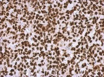
Immunohistochemical analysis of paraffin-embedded H1299 xenograft, using HMGA2(GTX100519) antibody at 1:500 dilution.
Antigen Retrieval: Trilogy™ (EDTA based, pH 8.0) buffer, 15min
Whole cell extract (30 μg) was separated by 15% SDS-PAGE, and the membrane was blotted with HMGA2 antibody (GTX100519) diluted at 1:10000. The HRP-conjugated anti-rabbit IgG antibody (GTX213110-01) was used to detect the primary antibody, and the signal was developed with Trident ECL plus-Enhanced.
Whole cell extract (30 μg) was separated by 15% SDS-PAGE, and the membrane was blotted with HMGA2 antibody - ChIP grade (GTX100519) diluted at 1:10000. The HRP-conjugated anti-rabbit IgG antibody (GTX213110-01) was used to detect the primary antibody.
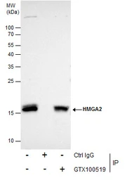
Immunoprecipitation of HMGA2 protein from HepG2 nuclear extracts using 5 μg of HMGA2 antibody (GTX100519).
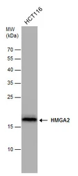
Western blot analysis was performed using HMGA2 antibody (GTX100519) diluted at 1:500.
EasyBlot anti-Rabbit IgG (GTX221666-01) was used as a secondary reagent.
HMGA2 antibody detects HMGA2 protein by western blot analysis. Whole cell extracts (30 μg) was separated by 15% SDS-PAGE, and the membrane was blotted with HMGA2 antibody (GTX100519) at a dilution of 1:10000. The HRP-conjugated anti-rabbit IgG antibody (GTX213110-01) was used to detect the primary antibody.
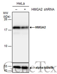
Non-transfected (–) and transfected (+) HeLa whole cell extracts (30 μg) were separated by 15% SDS-PAGE, and the membrane was blotted with HMGA2 antibody - ChIP grade (GTX100519) diluted at 1:50000. The HRP-conjugated anti-rabbit IgG antibody (GTX213110-01) was used to detect the primary antibody.
Immunohistochemical analysis of paraffin-embedded C2C12 xenograft, using HMGA2(GTX100519) antibody at 1:500 dilution.
Antigen Retrieval: Trilogy™ (EDTA based, pH 8.0) buffer, 15min
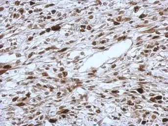
HMGA2 antibody - ChIP grade detects HMGA2 protein at nucleus in human HuCCT1 xenograft by immunohistochemical analysis.
Sample: Paraffin-embedded human HuCCT1 xenograft .
HMGA2 antibody - ChIP grade (GTX100519) diluted at 1:250.
Antigen Retrieval: Trilogy™ (EDTA based, pH 8.0) buffer, 15min
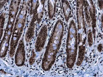
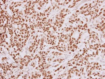
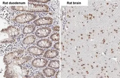
HMGA2 antibody detects HMGA2 protein at nucleus in rat duodenum by immunohistochemical analysis.
Sample: Paraffin-embedded rat duodenum.
HMGA2 antibody (GTX100519) diluted at 1:500.
Antigen Retrieval: Citrate buffer, pH 6.0, 15 min
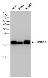
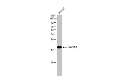
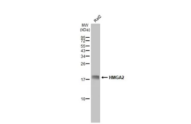
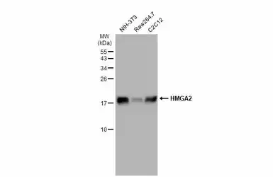
HMGA2 antibody detects HMGA2 protein by western blot analysis. Various whole cell extracts (30 μg) were separated by 15% SDS-PAGE, and the membrane was blotted with HMGA2 antibody (GTX100519) diluted at a dilution of 1:10000. The HRP-conjugated anti-rabbit IgG antibody (GTX213110-01) was used to detect the primary antibody.
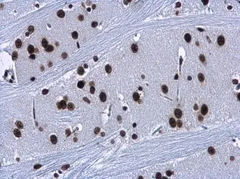
HMGA2 antibody detects HMGA2 protein at nucleus in rat brain by immunohistochemical analysis.
Sample: Paraffin-embedded rat brain.
HMGA2 antibody (GTX100519) diluted at 1:500.
Antigen Retrieval: Citrate buffer, pH 6.0, 15 min
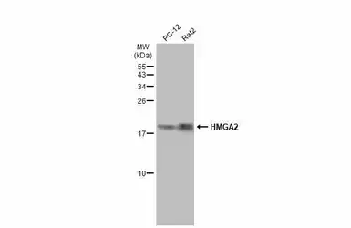
Various whole cell extracts (30 μg) were separated by 15% SDS-PAGE, and the membrane was blotted with HMGA2 antibody - ChIP grade (GTX100519) diluted at 1:1000. The HRP-conjugated anti-rabbit IgG antibody (GTX213110-01) was used to detect the primary antibody.
Various whole cell extracts (30 μg) were separated by 15% SDS-PAGE, and the membrane was blotted with HMGA2 antibody - ChIP grade (GTX100519) diluted at 1:1000. The HRP-conjugated anti-rabbit IgG antibody (GTX213110-01) was used to detect the primary antibody.
HMGA2 antibody - ChIP grade detects HMGA2 - ChIP grade protein by immunohistochemical analysis.Sample: Paraffin-embedded rat tissues.HMGA2 - ChIP grade stained by HMGA2 antibody - ChIP grade (GTX100519) diluted at 1:500.Antigen Retrieval: Citrate buffer, pH 6.0, 15 min
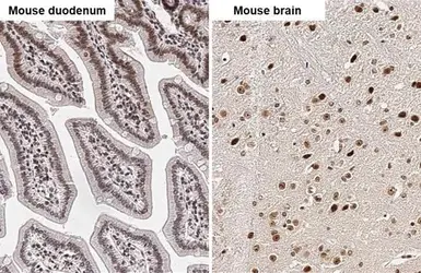
HMGA2 antibody - ChIP grade detects HMGA2 - ChIP grade protein by immunohistochemical analysis.Sample: Paraffin-embedded mouse tissues.HMGA2 - ChIP grade stained by HMGA2 antibody - ChIP grade (GTX100519) diluted at 1:500.Antigen Retrieval: Citrate buffer, pH 6.0, 15 min
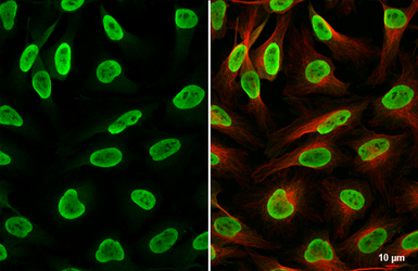
HMGA2 antibody - ChIP grade detects HMGA2 protein at nucleus by immunofluorescent analysis.Sample: HeLa cells were fixed in 4% paraformaldehyde at RT for 15 min.Green: HMGA2 stained by HMGA2 antibody - ChIP grade (GTX100519) diluted at 1:500.Red: alpha Tubulin, a cytoskeleton marker, stained by alpha Tubulin antibody [GT114] (GTX628802) diluted at 1:1000.
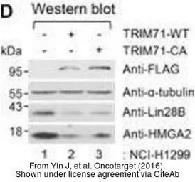
The data was published in the journal Oncotarget in 2016. PMID: 27821801
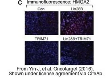
The data was published in the journal Oncotarget in 2016. PMID: 27821801

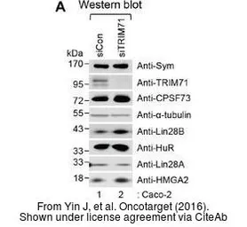
The data was published in the journal Oncotarget in 2016. PMID: 27821801
The data was published in the journal Oncotarget in 2016. PMID: 27821801
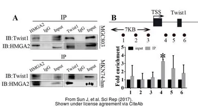
The data was published in the journal Sci Rep in 2017.PMID: 28533522