



G-Series Human Inflammation Array 3 Kit. Semi-quantitative measurement of 40 human cytokines. Suitable for all liquid sample types.
Human Inflammation Array GS3
Human Inflammation Array GS3
Contact Us for Bulk Request
Product Description
Specifications
| Size | 16 Sample Kit, 32 Sample Kit, 64 Sample Kit |
| Species | Human |
| Compatible Sample Types | Cell Culture Supernatants, Plasma, Serum, Tissue Lysates, Cell Lysates |
| Quantitative/Semi-Quantitative | Semi-Quantitative |
| Number of Targets Detected | 40 |
| Solid Support | Glass Slide |
| Design Principle | Sandwich-based |
| Method Of Detection | Fluorescence Laser Scanner |
| Research Area | Inflammation |
| Shipping Type | Blue ice |
| Storage | -20°C |
Introduction
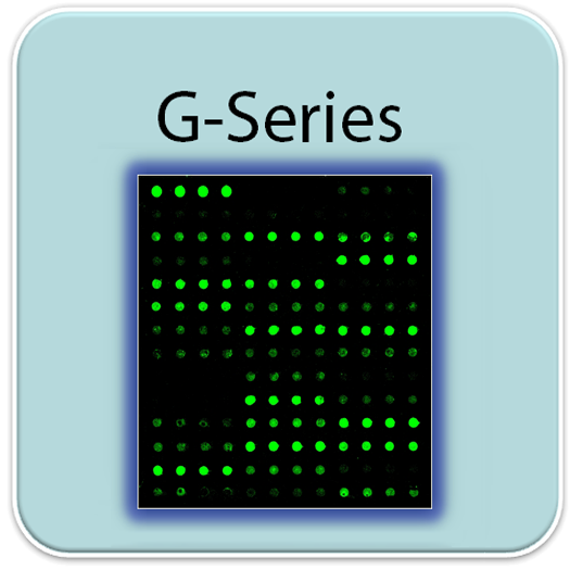
RayBio® G-Series Arrays are glass slide-based antibody arrays which allow researchers to conduct rapid, accurate expression profiling of hundreds of cytokines, chemokines, growth factors, proteases, soluble receptors and other proteins from any biological fluid. Like a traditional sandwich-based ELISA, this array uses a matched pair of cytokine-specific antibodies for detection. Like the Quantibody® Multiplex ELISA Arrays, G-Series utilizes a highly sensitive and stable fluorescent readout which can be detected by most laser fluorescent scanner systems (see below for more info). After capturing the spot densities with a laser scanner, normalization of the raw data can be easily calculated by the researcher, or by a quick copy-paste into our excel-based Analysis Tool software.
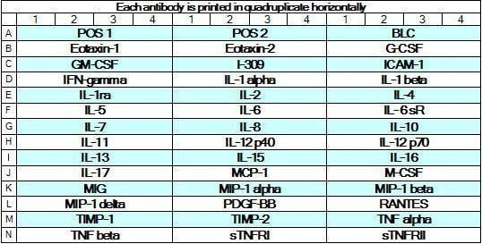
This array as well as all catalog numbers beginning with 'GS' differ from the classic G-Series Arrays in a few important ways. First, each capture antibody is printed in quadruplicate instead of duplicate, delivering higher precision. Secondly, this array features the same antibody panels used in our Quantibody Arrays, allowing a seamless transition to our quantitative multiplex assay platform. Lastly, all 16 wells are spotted as sub-arrays, delivering easy handling of 16 samples simultaneously while consuming low sample volumes (10 - 100 µl per array).
This array as well as all catalog numbers beginning with 'GS' differ from the classic G-Series Arrays in a few important ways. First, each capture antibody is printed in quadruplicate instead of duplicate, delivering higher precision. Secondly, this array features the same antibody panels used in our Quantibody Arrays, allowing a seamless transition to our quantitative multiplex assay platform. Lastly, all 16 wells are spotted as sub-arrays, delivering easy handling of 16 samples simultaneously while consuming low sample volumes (10 - 100 µl per array).
Product Features
- High sensitivity (pg/mL to ng/mL levels)
- High specificity and system reproducibility
- Suitable for diverse sample types
- Low sample volume requirement: 50 µL or less
- Get results same day (6-hour processing time)
- Well-suited for high throughput assays
- Test 16 samples on each slide
- Data analysis software provides one-step computation
Target Names
| Scroll over each target protein for more information | ||||
|---|---|---|---|---|
BLC (CXCL13)
| Eotaxin-1 (CCL11)
| Eotaxin-2 (MPIF-2/CCL24)
| GCSF
| GM-CSF
|
I-309 (TCA-3/CCL1)
| ICAM-1 (CD54)
| IFN-gamma
| IL-1 alpha (IL-1 F1)
| IL-1 beta (IL-1 F2)
|
IL-1 ra (IL-1 F3)
| IL-2
| IL-4
| IL-5
| IL-6
|
IL-6 R
| IL-7
| IL-8 (CXCL8)
| IL-10
| IL-11
|
IL-12 p40
| IL-12 p70
| IL-13
| IL-15
| IL-16
|
IL-17A
| MCP-1 (CCL2)
| M-CSF
| MIG (CXCL9)
| MIP-1 alpha (CCL3)
|
MIP-1 beta (CCL4)
| MIP-1 delta (CCL15)
| PDGF-BB
| RANTES (CCL5)
| TIMP-1
|
TIMP-2
| TNF alpha
| TNF beta (TNFSF1B)
| TNF RI (TNFRSF1A)
| TNF RII (TNFRSF1B)
|
Application Notes
Suggested Application
- Multiplexed Protein Detection
- Detection of Relative Protein Expression
- Detecting Patterns of Cytokine Expression
- Biomarker Screening
- Identifying Key Factors
- Confirming a Biological Process
Kit Components
- Antibody Array Slide/s
- Blocking Buffer
- Wash Buffer 1
- Wash Buffer 2
- Biotinylated Detection Antibody Cocktail
- Streptavidin-Conjugated Fluor
- 30 ml Centrifuge Tube
- Adhesive Plastic Strips
- Manual
Other Materials Required
- Distilled or deionized water
- Small plastic boxes or containers
- Pipettors, pipette tips and other common lab consumables
- Orbital shaker or oscillating rocker
- Aluminum foil
- Gene microarray scanner or similar laser fluorescence scanner
View Compatible Laser Scanners
Don't have a compatible scanner? RayBiotech now offers FREE scanning service for all RayBio glass slide antibody arrays! Learn More
Protocol Outline
- Dry the glass slide
- Prepare Standards
- Block array surface
- Incubate with samples
- Incubate with Biotinylated Detection Antibody Cocktail
- Incubate with Streptavidin-Conjugated Fluor
- Disassemble the glass slide
- Scan with a gene microarray laser scanner
- Perform densitometry and analysis
Storage/Stability
For best results, store the entire kit frozen at -20°C upon arrival. Stored frozen, the kit will be stable for at least 6 months which is the duration of the product warranty period. Once thawed, store array slide(s), detection antibody cocktail, and Cy3-Conjugated Streptavidin at -20°C and all other reagents undiluted at 4°C for no more than 3 months.
documents
GAL (GAL-QAH-INF-3.gal)
Excel (GSH-INF-3_Analysis_Tool.xlsx)
citations
Identification of a small-molecule targeting PLAGL2 DNA-binding domain inhibits extracellular matrix formation and enhances lenvatinib sensitivity in hepatocellular carcinoma
- Species:
- Human
- Sample Type:
- Serum (serum-free medium of Huh-7 cells)
- Year:
- 2026
- Journal Name:
- Acta Pharmaceutica Sinica B
- PubMed ID:
- 41909729
Plasmatic CXCL13 as a biomarker of primary resistance to immunotherapy and impaired peripheral immunity in non-small cell lung cancer
- Species:
- Human
- Sample Type:
- Plasma (baseline and post-first-cycle immunotherapy)
- Year:
- 2026
- Journal Name:
- Biomarker Research
- PubMed ID:
- 42163420
PFDN5 synergizes with anti-PD1 therapy to promote triple-negative breast cancer cell death through JAK2/STAT3/c-Myc signaling axis
- Species:
- Human
- Sample Type:
- Cell Lysate (cell lysates of TNBC cells); Tissue Lysate / Homogenate
- Year:
- 2026
- Journal Name:
- Journal of Translational Medicine
- PubMed ID:
- 42032729
hAECs restore follicular development in premature ovarian insufficiency via IGFBP2/IGF1R-mediated intercellular communication
- Species:
- Human
- Sample Type:
- Cell Culture Supernatant (concentrated hAEC conditioned medium)
- Year:
- 2026
- Journal Name:
- Stem Cell Research & Therapy
- PubMed ID:
- 42144654
Lysosome-mediated aggregation of galactose-deficient IgA1 with transferrin receptor 1 links to IgA nephropathy
- Species:
- Human
- Sample Type:
- Cell Culture Supernatant (conditioned medium from mesangial cells)
- Year:
- 2025
- Journal Name:
- Nature Communications
- PubMed ID:
- 40593574
Novel full-thickness biomimetic corneal model for studying pathogenesis and treatment of diabetic keratopathy
- Species:
- Human
- Sample Type:
- Cell Culture Supernatant (full-thickness corneal model)
- Year:
- 2025
- Journal Name:
- Materials Today Bio
- PubMed ID:
- 39807180
Elevated LIF and JAK-STAT activation drive severe COVID-19 in myeloma patients receiving the BCMA-CD3 bispecific antibody Elranatamab
- Species:
- Human
- Sample Type:
- Plasma (myeloma patients with COVID-19)
- Year:
- 2025
- Journal Name:
- Journal of Translational Medicine
- PubMed ID:
- 39865264
Immunoregulatory role of the gut microbiota in inflammatory depression
- Species:
- Human
- Sample Type:
- Other / Unspecified (intestinal mucosal samples)
- Year:
- 2024
- Journal Name:
- Nature Communications
- PubMed ID:
- 38589368
Single-cell transcriptomics and epigenomics unravel the role of monocytes in neuroblastoma bone marrow metastasis
- Species:
- Human
- Sample Type:
- CLB-Ma cell, CHLA90 cell, peripheral blood mononuclear cells (PBMCs)
- Year:
- 2023
- Journal Name:
- Nature Communications
IL-1β-associated NNT acetylation orchestrates iron-sulfur cluster maintenance and cancer immunotherapy resistance
- Species:
- Human
- Sample Type:
- gastric tissue
- Year:
- 2023
- Journal Name:
- Molecular Cell