Mouse H-2Db Antibody : LE/AF

Référence OASB00710
Conditionnement : 0.5mg
Marque : Aviva Systems Biology
Contactez votre distributeur local :
Mouse H-2Db Antibody : LE/AF (OASB00710)
| Datasheets/Manuals | Printable datasheet for Mouse H-2Db Antibody : LE/AF (OASB00710) |
|---|
| Tested Species Reactivity | Mouse |
|---|---|
| Predicted Species Reactivity | Mouse |
| Clonality | Monoclonal |
| Clone | 28-14-8 |
| Isotype | IgG2a |
| Host | Mouse |
| Conjugation | LE: Low Endotoxin, AF: Azide Free |
| Application | FC, IHC-F, IP, BL, CMCD |
| Additional Information | Description: The "classical" MHC Class I molecules are histocompatibility antigens encoded by the H-2 gene complex and consist of heterodimers of highly polymorphic alpha chains noncovalently associated with the invariant beta2-microglobulin. These antigens are expressed on most nucleated cells but expression varies on different cell types. MHC Class I molecules present endogenously synthesized peptides to CD8+ T lymphocytes which are usually cytotoxic T cells. MHC Class I antigens expressed on thymic epithelial cells regulate the positive and negative selection of CD8+ T cells during T cell ontogeny. The monoclonal antibody 28-14-8 binds to the alpha3 domain of H-2Db in the presence or absence of beta2 microglobulin. It cross reacts with the alpha3 domain of H-2Ld, but not Kd or Dd, and with H-2Dq and/or Lq. The antibody has been shown to block H-2Ld-specific and H-2Ld-restricted antigen-specific lysis of target cells by cytotoxic T lymphocytes but it does not block recognition of H-2Ld-positive target cells by Ly-6G2-positive NK cells. |
| Reconstitution and Storage | Store at -20C |
| Immunogen | C3H.SW mouse splenocytes |
| Concentration | 0.5 mg/mL |
| Specificity | H-2Db |
| Characterization | To ensure lot- to- lot consistency, each batch of product is tested by flow cytometry to conform tocharacteristics of a standard reference reagent. |
| Warning | Reagents contain sodium azide. Sodium azide is very toxic if ingested or inhaled. Avoid contact with skin,eyes, or clothing. Wear eye or face protection when handling. If skin or eye contact occurs, wash withcopious amounts of water. If ingested or inhaled, contact a physician immediately. Sodium azide yieldstoxic hydrazoic acid under acidic conditions. Dilute azide- containing compounds in running water beforediscarding to avoid accumulation of potentially explosive deposits in lead or copper plumbing. |
| Dilution | Flow Cytometry: Purified antibody Fluorescein conjugate Biotin conjugate R-phycoerythrin conjugate <=1 ug/106 cells <=1 ug/106 cells <=1 ug/106 cells <=0.5 ug/106 cells |
| Application Info | Flow cytometry13, Complement-dependent cytotoxicity1, 2, 9, Immunoprecipitation8 |
| Other Applications Data | Since applications vary, you should determine the optimum workingdilution of the product that is appropriate for your specific need. |
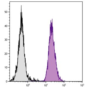
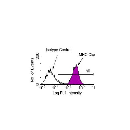
| Other Applications Image 1 Data | Amount Used: 1 Ug/106 cells Splenocytes from DBA1/J mice were stained with mouse anti-mouse H-2Db-FITC. Small lymphocytes were then gated and analyzed on a FACScan flow cytometer (BDIS, San Jose, CA). |
| Storage | - The purified (UNLB) antibody is supplied as 0.5 mg of purified immunoglobulin in 1.0 mL of 100 mMborate buffered saline, pH 8.2. No preservatives or amine- containing buffer salts added. Store at 2- 8 C - The fluorescein (FITC) conjugate is supplied as 0.5 mg in 1.0 mL of PBS/NaN3. Store at 2- 8 C - The biotin (BIOT) conjugate is supplied as 0.5 mg in 1.0 mL of PBS/NaN3. Store at 2- 8 C - The R- phycoerythrin (R- PE) conjugate is supplied as 0.1 mg in 1.0 mL of PBS/NaN3 and a stabilizingagent. Store at 2- 8 C. Do not freeze! - The low endotoxin, azide- free (LE/AF) antibody is supplied as 0.5 mg purified immunoglobulin in 1.0 mLof PBS. Aliquot and store at or below - 20 C - Protect conjugated forms from light. Aliquot and freeze the low endotoxin, azide- free product at - 20 Cimmediately upon receipt. Each reagent is stable for the period shown on the bottle label if stored asdirected. |
| Reference | 1. Ozato, K., T.H. Hansen, and D.H. Sachs. 1980. J. Immunol. 125:2473 2. Ozato, K., and D.H. Sachs. 1981. J. Immunol. 126:317 3. Lawlor, D.A., et al. 1990. Annu. Rev. Immunol. 8:23 4. Bjorkman, P.J., et al. 1987. Nature 329:506 5. Yewdell, J.W., and J.R. Bennett. 1990. Cell 62:203 6. Zijlstra, M., et al. 1990. Nature 344:742 7. Allen, H., D. Wraith, P. Pala, B. Askonas, and R.A. Flavell. 1984. Nature 309:279 8. Allen, H, J. Fraser, D. Flyer, S. Calvin, and R. Flavell. 1986. Proc. Natl. Acad. Sci. USA 83:74479. Woodward, J.G., A. Orn, R.C. Harmon, R.S. Goodenow, L. Hood, and J.A. Frelinger. 1982. Proc. Natl. Acad. Sci. USA 79:3613 10. Orn, A., R.S. Goodenow, L. Hood, P.R. Brayton, J.G. Woodward, R.C. Harmon, and J.A. Frelinger. 1982. Nature 297:415 11. Kundig, T.M., M.F. Bachmann, C. DiPaolo, et al. 1995. Science 268:1343 12. Mason, L.H., J.R. Ortaldo, H.A. Young, V. Kumar, M. Bennett, and S.K. Anderson. 1995. J. Exp. Med. 182:293 13. Gri,G., C. Chiodoni, E. Gallo, A. Stoppacciaro, F.Y. Liew, and M.P. Colombo. 2002. Cancer Res. 62:4390. |
|---|---|
| Gene Symbol | H2-D1 |
| Gene Full Name | H-2 class I histocompatibility antigen, D-B alpha chain |
| Alias Symbols | H-2D, H2-D, H2-K1 |
| NCBI Gene Id | 14964 |
| Protein Name | H-2 class I histocompatibility antigen, D-B alpha chain |
| Uniprot ID | P01899 |
| Protein Accession # | NP_034510.3 |
| Nucleotide Accession # | NM_010380.3 |