

RIPA Lysis Buffer

Référence MBS1753872-5x100mL
Conditionnement : 5x100mL
Marque : MyBiosource
Contactez votre distributeur local :

Téléphone : +1 850 650 7790
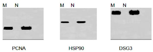
Example Data using RIPA Lysis Buffer:
M: Hela Cells N: A549 Cells
Total proteins (20ug) lysed using RIPA Lysis Buffer were separated by SDS-PAGE and transferred to a nitrocellulose membrane. Incubate with primary antibodies. And Boster ECL substrate was used to generate the images.
M: Hela Cells N: A549 Cells
Total proteins (20ug) lysed using RIPA Lysis Buffer were separated by SDS-PAGE and transferred to a nitrocellulose membrane. Incubate with primary antibodies. And Boster ECL substrate was used to generate the images.
RIPA Reagent – Assistive Reagent
For Research Use Only. Not for use in diagnostic procedures. Catalog #: MBS1753872Description: RIPA Lysis Buffer is a complete cell lysis solution reagent used for rapid and efficient total cell lysis and solubilization of proteins from both adherent and suspension cultured mammalian cells, effectively extracting cytoplasmic, nuclear and… More details
Product Overview
Online Datasheet in PDFProduct Name
RIPA, Reagent
Full Product Name
RIPA Lysis Buffer
Form/Format
Liquid. 50mM Tris-HCl pH7.6, 150mM NaCl, 1% NP-40, 0.5% sodium deoxycholate, 0.1% SDS
Concentration
Recommended working concentration
10mL RIPA Lysis Buffer per gram of tissue
0.5mL RIPA Lysis Buffer per 5.0x10^6 cells in suspension
0.5mL RIPA Lysis Buffer per 5.0x10^6 adherent mammalian cells (lot specific)
10mL RIPA Lysis Buffer per gram of tissue
0.5mL RIPA Lysis Buffer per 5.0x10^6 cells in suspension
0.5mL RIPA Lysis Buffer per 5.0x10^6 adherent mammalian cells (lot specific)
Preparation and Storage
Upon receipt store at 4 degree C. RIPA Lysis Buffer is stable for one year. Product is shipped on ice.
Compatibility with reagents
Fully compatible with Broad Spectrum Protease Inhibitor Cocktail and Broad Spectrum Phosphatase Inhibitor Cocktail
Reagent Type
Western Blotting related reagent; Cell lysis buffer; Universal tissue extraction buffer; Detergent solution
Usage
Extraction of cytoplasmic, membrane and nuclear proteins
Important Product Information
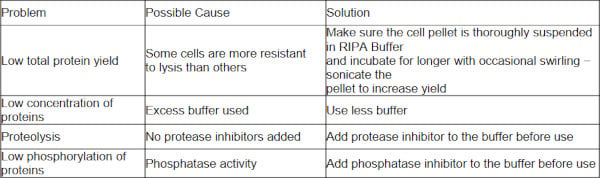
If desired, add protease inhibitor and phosphatase inhibitor to the lysis buffer to prevent proteolysis and maintain phosphorylation status of proteins.
Some protein kinases and other enzymes may be sensitive to the components of the RIPA Lysis Buffer, resulting in their decreased activity. In such cases, prepare a RIPA Lysis Buffer that does not contain sodium deoxycholate and SDS.
Some protein kinases and other enzymes may be sensitive to the components of the RIPA Lysis Buffer, resulting in their decreased activity. In such cases, prepare a RIPA Lysis Buffer that does not contain sodium deoxycholate and SDS.
Additional Materials Required
Protease inhibitor and phosphatase inhibitor
2mL microcentrifuge tubes
Tissue homogenizer
Microcentrifuge capable of spinning at 10,000 xg
Cell scraper
2mL microcentrifuge tubes
Tissue homogenizer
Microcentrifuge capable of spinning at 10,000 xg
Cell scraper
Procedure for Lysis of Monolayer-cultured Adherent Mammalian Cells
Note: Pre-chill an appropriate volume of RIPA Lysis Buffer at 4 degree C. If desired, add protease inhibitor and phosphatase inhibitor to the lysis buffer immediately before use.
1. In a microcentrifuge tube, resuspend 5x10^6 cells in the growth media by scraping the cells off the surface of the plate with a cell scraper. Centrifuge harvested cell suspension at 600xg for 5min, then carefully remove and discard the supernatant.
2. Resuspend the cells in chilled PBS. Centrifuge at 600xg for 5min, then carefully remove and discard the supernatant.
3. Add 0. 5mL of chilled RIPA lysis buffer to the cell pellet. Vortex briefly. Incubate on ice for 30 minutes.
4. Centrifuge samples at 14000xg for 10 minutes.
5. Transfer supernatant to a new tube for further analysis.
Note: RIPA lysis buffer can be added directly to the flask containing cells. Please see the following procedures.
1. Carefully remove culture medium from adherent cells.
2. Wash cells with chilled PBS. Carefully remove PBS.
3. Add chilled RIPA lysis buffer to the cells. Vortex briefly. Incubate on ice for 30 minutes. (For the volume of the lysis buffer, follow the instructions listed below)
SIZE of the plate/surface area: Volume of the lysis buffer
100mm: 500-1000uL
60mm: 250-500uL
6-well plate: 200-400uL per well
-well plate: 100-200uL per well
96-well plate: 50-100uL per well
4. Centrifuge samples at 14000xg for 10 minutes.
5. Transfer supernatant to a new tube for further analysis.
1. In a microcentrifuge tube, resuspend 5x10^6 cells in the growth media by scraping the cells off the surface of the plate with a cell scraper. Centrifuge harvested cell suspension at 600xg for 5min, then carefully remove and discard the supernatant.
2. Resuspend the cells in chilled PBS. Centrifuge at 600xg for 5min, then carefully remove and discard the supernatant.
3. Add 0. 5mL of chilled RIPA lysis buffer to the cell pellet. Vortex briefly. Incubate on ice for 30 minutes.
4. Centrifuge samples at 14000xg for 10 minutes.
5. Transfer supernatant to a new tube for further analysis.
Note: RIPA lysis buffer can be added directly to the flask containing cells. Please see the following procedures.
1. Carefully remove culture medium from adherent cells.
2. Wash cells with chilled PBS. Carefully remove PBS.
3. Add chilled RIPA lysis buffer to the cells. Vortex briefly. Incubate on ice for 30 minutes. (For the volume of the lysis buffer, follow the instructions listed below)
SIZE of the plate/surface area: Volume of the lysis buffer
100mm: 500-1000uL
60mm: 250-500uL
6-well plate: 200-400uL per well
-well plate: 100-200uL per well
96-well plate: 50-100uL per well
4. Centrifuge samples at 14000xg for 10 minutes.
5. Transfer supernatant to a new tube for further analysis.
Procedure for Lysis of Suspension-cultured Mammalian Cells
Note: Pre-chill an appropriate volume of RIPA Lysis Buffer at 4 degree C. If desired, add protease inhibitor and phosphatase inhibitor to the lysis buffer immediately before use.
1. In a microcentrifuge tube, harvest 5x10⁶ cells by centrifugation at 600xg for 5min. Carefully remove and discard the supernatant.
2. Resuspend the cells in chilled PBS. Centrifuge at 600xg for 5min, then carefully remove and discard the supernatant.
3. Add 0. 5mL of chilled RIPA lysis buffer to the cell pellet. Vortex briefly. Incubate on ice for 30 minutes.
4. Centrifuge samples at 14000xg for 10 minutes.
5. Transfer supernatant to a new tube for further analysis.Procedure for Lysis of Tissues:
Note: Pre-chill an appropriate volume of RIPA Lysis Buffer at 4 degree C. If desired, add protease inhibitor and phosphatase inhibitor to the lysis buffer immediately before use.
1. Place the fresh tissue into chilled PBS and rinse several times. Mince the tissue into small pieces.
2. Add RIPA Lysis Buffer to the tissue at 10:1. (i.e. Add 10mL cilled lysis buffer per gram of tissue.) Use a smaller volume of reagent if a more concentrated protein extract is required.
3. Homogenize for several minutes at high speed until no tissue chunks remain.
4. Incubate on ice for 30 minutes.
5. Centrifuge at ~10000 x g for 10 minutes.
6. Transfer supernatant to a new tube for further analysis.
1. In a microcentrifuge tube, harvest 5x10⁶ cells by centrifugation at 600xg for 5min. Carefully remove and discard the supernatant.
2. Resuspend the cells in chilled PBS. Centrifuge at 600xg for 5min, then carefully remove and discard the supernatant.
3. Add 0. 5mL of chilled RIPA lysis buffer to the cell pellet. Vortex briefly. Incubate on ice for 30 minutes.
4. Centrifuge samples at 14000xg for 10 minutes.
5. Transfer supernatant to a new tube for further analysis.Procedure for Lysis of Tissues:
Note: Pre-chill an appropriate volume of RIPA Lysis Buffer at 4 degree C. If desired, add protease inhibitor and phosphatase inhibitor to the lysis buffer immediately before use.
1. Place the fresh tissue into chilled PBS and rinse several times. Mince the tissue into small pieces.
2. Add RIPA Lysis Buffer to the tissue at 10:1. (i.e. Add 10mL cilled lysis buffer per gram of tissue.) Use a smaller volume of reagent if a more concentrated protein extract is required.
3. Homogenize for several minutes at high speed until no tissue chunks remain.
4. Incubate on ice for 30 minutes.
5. Centrifuge at ~10000 x g for 10 minutes.
6. Transfer supernatant to a new tube for further analysis.
Precautions
All steps of protein lysis should be operated on ice or at 4 degree C.
Use BCA Protein Assay kit to quantify lysed proteins. Bradford Protein Assay kit is not recommended.
There might be some transparent gel complex containing genomic DNA in lysed proteins. The protein fractions can be used for further analysis after centrifugation if target proteins have little connection with genomic DNA. When detecting target proteins related closely to genomic DNA, sonicate gel complex and then centrifuge to collect supernatant for further analysis. Common transcription factors such as NFKB, p53 can be detected without sonication.
Use BCA Protein Assay kit to quantify lysed proteins. Bradford Protein Assay kit is not recommended.
There might be some transparent gel complex containing genomic DNA in lysed proteins. The protein fractions can be used for further analysis after centrifugation if target proteins have little connection with genomic DNA. When detecting target proteins related closely to genomic DNA, sonicate gel complex and then centrifuge to collect supernatant for further analysis. Common transcription factors such as NFKB, p53 can be detected without sonication.
Product Categories/Family for RIPA reagent
Assistive Reagent
ISO Certification
Manufactured in an ISO 13485:2003 and EN ISO 13485:2012 Certified Laboratory.
Testing Data (TD) of RIPA reagent
Example Data using RIPA Lysis Buffer:
M: Hela Cells N: A549 Cells
Total proteins (20ug) lysed using RIPA Lysis Buffer were separated by SDS-PAGE and transferred to a nitrocellulose membrane. Incubate with primary antibodies. And Boster ECL substrate was used to generate the images.
M: Hela Cells N: A549 Cells
Total proteins (20ug) lysed using RIPA Lysis Buffer were separated by SDS-PAGE and transferred to a nitrocellulose membrane. Incubate with primary antibodies. And Boster ECL substrate was used to generate the images.
Other Notes
Small volumes of RIPA reagent vial(s) may occasionally become entrapped in the seal of the product vial during shipment and storage. If necessary, briefly centrifuge the vial on a tabletop centrifuge to dislodge any liquid in the container`s cap. Certain products may require to ship with dry ice and additional dry ice fee may apply.
Searchable Terms for RIPA purchase
MBS1753872 is a reagent targeting RIPA. Also known as RIPA Lysis Buffer.
Additional Details
NCBI/Uniprot data below describe general gene information for RIPA. It may not necessarily be applicable to this product.
Protein Family
HTH-type transcriptional regulator
Precautions
All of MyBioSource's Products are for scientific laboratory research purposes and are not for diagnostic, therapeutics, prophylactic or in vivo use. Through your purchase, you expressly represent and warrant to MyBioSource that you will properly test and use any Products purchased from MyBioSource in accordance with industry standards. MyBioSource and its authorized distributors reserve the right to refuse to process any order where we reasonably believe that the intended use will fall outside of our acceptable guidelines.
Disclaimer
While every efforts were made to ensure the accuracy of the information provided in this datasheet, MyBioSource will not be liable for any omissions or errors contained herein. MyBioSource reserves the right to make changes to this datasheet at any time without prior notice.
It is the responsibility of the customer to report product performance issues to MyBioSource within 30 days of receipt of the product. Please visit our Terms & Conditions page for more information.
It is the responsibility of the customer to report product performance issues to MyBioSource within 30 days of receipt of the product. Please visit our Terms & Conditions page for more information.
Product Note
Select online data sheet information is drawn from bioinformatics databases, occasionally resulting in ambiguous or non-relevant product information. It is the responsibility of the customer to review, verify, and evaluate the information to make sure it matches their requirements before purchasing the kit. Our ELISA Kit assays are dynamic research tools and sometimes they may be updated and improved. If the format of this assay is important to you then please request the current manual or contact our technical support team with a presales inquiry before placing an order. We will confirm the current details of the assay. We cannot guarantee the sample manual posted online is the most current manual, it is intended to serve as an example only. Please refer to the instructions For Use provided with the assay kit for precise details.