



Immunofluorescence
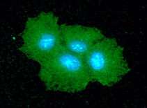
Immunofluorescence analysis of SAR1B in Hep3B cells. The cell was stained with SAR1B monoclonal antibody, clone AT1C7 (Cat # MAB11207) (1:100). The secondary antibody (green) was used Alexa Fluor 488. DAPI was stained the cell nucleus (blue).
Flow Cytometry
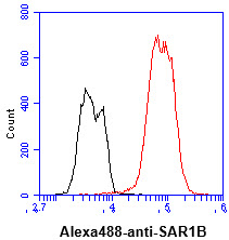
Flow cytometry analysis of SAR1B in Hep3B cell line, staining at 2-5 ug for 1x10⁶ cells (red line). The secondary antibody used goat anti-mouse IgG Alexa fluor 488 conjugate. Isotype control antibody was mouse IgG (black line).
Western Blot (Tissue lysate)
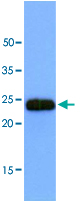
Western blot analysis of mouse muscle extract (40 ug) by using SAR1B monoclonal antibody, clone AT1C7 (Cat # MAB11207) (1:1000). Proteins were visualized using a goat anti-mouse secondary antibody conjugated to HRP and an ECL detection system.
- Specifications
Product Description
Mouse monoclonal antibody raised against partial recombinant SAR1B.
Immunogen
Recombinant protein corresponding to amino acids 1-198 of human SAR1B.
Host
Mouse
Reactivity
Human
Form
Liquid
Purification
Protein A purification
Concentration
1 mg/mL
Isotype
IgG1, kappa
Recommend Usage
ELISA
Flow Cytometry
Immunocytochemistry
Immunofluorescence
Western Blot
The optimal working dilution should be determined by the end user.Storage Buffer
In PBS, pH 7.4 (10% glycerol, 0.02% sodium azide).
Storage Instruction
Store at 2°C to 8°C for 1 week. For long term storage, aliquot and store at -20°C to -80°C.
Aliquot to avoid repeated freezing and thawing. - Applications
Immunofluorescence
Immunofluorescence analysis of SAR1B in Hep3B cells. The cell was stained with SAR1B monoclonal antibody, clone AT1C7 (Cat # MAB11207) (1:100). The secondary antibody (green) was used Alexa Fluor 488. DAPI was stained the cell nucleus (blue).Immunocytochemistry
Flow Cytometry
Flow cytometry analysis of SAR1B in Hep3B cell line, staining at 2-5 ug for 1x10⁶ cells (red line). The secondary antibody used goat anti-mouse IgG Alexa fluor 488 conjugate. Isotype control antibody was mouse IgG (black line).Enzyme-linked Immunoabsorbent Assay
Western Blot (Tissue lysate)
Western blot analysis of mouse muscle extract (40 ug) by using SAR1B monoclonal antibody, clone AT1C7 (Cat # MAB11207) (1:1000). Proteins were visualized using a goat anti-mouse secondary antibody conjugated to HRP and an ECL detection system. - Gene Info — SAR1B
- Interactomes