



Immunofluorescence
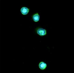
Immunofluorescent staining of THP-1 cells line. The cell was stained with TLR7 monoclonal antibody, clone 4F4 (Cat # MAB1062) (1:100). The secondary antibody (green) was used Alexa Fluor 488. DAPI was stained the cell nucleus (blue).
Flow Cytometry
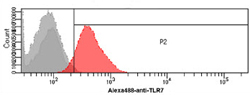
Flow cytometry analysis of THP-1 cells. The cell was stained with TLR7 monoclonal antibody, clone 4F4 (Cat # MAB1062) at 2-5 ug for 1x10⁶ cells (red). A goat anti mouse IgG (Alexa fluor 488) was used as the secondary antibody. Mouse monoclonal IgG was used as the isotype control (dark gray), cells without incubation with primary and secondary antibody was used as the negative control (light gray).
- Specifications
Product Description
Mouse monoclonal antibody raised against partial recombinant TLR7.
Immunogen
Recombinant protein corresponding to amino acids 451-500 of human TLR7.
Host
Mouse
Reactivity
Human
Form
Liquid
Purification
Protein G affinity chromatography
Isotype
IgG1, kappa
Recommend Usage
ELISA
Flow Cytometry
Immunocytochemistry (1:100)
Immunofluorescence (1:100)
The optimal working dilution should be determined by the end user.Storage Buffer
In PBS, pH 7.4 (10% glycerol, 0.02% sodium azide).
Storage Instruction
Store at 2°C to 8°C for 1 week. For long term storage, aliquot and store at -20°C to -80°C.
Aliquot to avoid repeated freezing and thawing. - Applications
Immunofluorescence
Immunofluorescent staining of THP-1 cells line. The cell was stained with TLR7 monoclonal antibody, clone 4F4 (Cat # MAB1062) (1:100). The secondary antibody (green) was used Alexa Fluor 488. DAPI was stained the cell nucleus (blue).Immunocytochemistry
Flow Cytometry
Flow cytometry analysis of THP-1 cells. The cell was stained with TLR7 monoclonal antibody, clone 4F4 (Cat # MAB1062) at 2-5 ug for 1x10⁶ cells (red). A goat anti mouse IgG (Alexa fluor 488) was used as the secondary antibody. Mouse monoclonal IgG was used as the isotype control (dark gray), cells without incubation with primary and secondary antibody was used as the negative control (light gray).Enzyme-linked Immunoabsorbent Assay
- Gene Info — TLR7
Entrez GeneID
51284GeneBank Accession#
NM_016562Protein Accession#
Q9NYK1Gene Name
TLR7
Gene Alias
-
Gene Description
toll-like receptor 7
Omim ID
300365Gene Ontology
HyperlinkGene Summary
The protein encoded by this gene is a member of the Toll-like receptor (TLR) family which plays a fundamental role in pathogen recognition and activation of innate immunity. TLRs are highly conserved from Drosophila to humans and share structural and functional similarities. They recognize pathogen-associated molecular patterns (PAMPs) that are expressed on infectious agents, and mediate the production of cytokines necessary for the development of effective immunity. The various TLRs exhibit different patterns of expression. This gene is predominantly expressed in lung, placenta, and spleen, and lies in close proximity to another family member, TLR8, on chromosome X. [provided by RefSeq
Other Designations
OTTHUMP00000022919
- Interactomes
- Pathways
- Diseases
- Publication Reference
- TLR7: A new sensor of viral infection.
Crozat K, Beutler B.
PNAS 2004 May; 101(18):6835.
Application:Flow Cyt, Human, Human mammalian cells.
- Molecular basis for the immunostimulatory activity of guanine nucleoside analogs: activation of Toll-like receptor 7.
Lee J, Chuang TH, Redecke V, She L, Pitha PM, Carson DA, Raz E, Cottam HB.
PNAS 2003 May; 100(11):6646.
- The adaptor molecule TIRAP provides signalling specificity for Toll-like receptors.
Horng T, Barton GM, Flavell RA, Medzhitov R.
Nature 2002 Nov; 420(6913):329.
- TLR7: A new sensor of viral infection.
- Literature Request
Cancer Bioreagents