

Acridine Orange

Cat# MBS258066-0,5mL
Size : 0.5mL
Brand : MyBiosource
Contact local distributor :

Phone : +1 850 650 7790
ACRIDINE ORANGE STAINING OF JURKAT CELLS
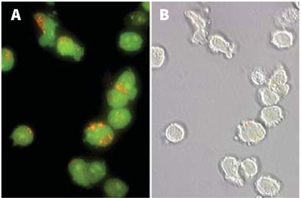
Normal Jurkat cells stained with Acridine Orange (AO) show orange lysosomal staining. Jurkat cells were stained with 5uM AO in PBS for 60 minutes at 37°C. Photomicrographs were taken using a Nikon Eclipse E800 photomicroscope using a 460-500 nm excitation filter and a 505-560 nm emission / barrier filter set at 300X. Photo B shows the corresponding DIC image of the cells in A (AO appears faintly).
Normal Jurkat cells stained with Acridine Orange (AO) show orange lysosomal staining. Jurkat cells were stained with 5uM AO in PBS for 60 minutes at 37°C. Photomicrographs were taken using a Nikon Eclipse E800 photomicroscope using a 460-500 nm excitation filter and a 505-560 nm emission / barrier filter set at 300X. Photo B shows the corresponding DIC image of the cells in A (AO appears faintly).
ACRIDINE ORANGE STAINING OF MCF-7 CELLS
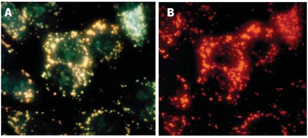
Apoptosis was induced in human breast cancer MCF-7 cells by treating them with 0.15 uM camptothecin for 24 hours at 37°C. Cells were stained with AO in PBS for 30 minutes, then washed twice in PBS. Cells were photographed with a Nikon Microphot-FXA epifluorescence microscope at 40X using either a blue light excitation (492 nm) with a 540-550 nm emission filter (A, lysosomes appear yellowish green), or green light excitation (540 nm) with a long pass >640 nm barrier filter (B, lysosomes appear red).
Apoptosis was induced in human breast cancer MCF-7 cells by treating them with 0.15 uM camptothecin for 24 hours at 37°C. Cells were stained with AO in PBS for 30 minutes, then washed twice in PBS. Cells were photographed with a Nikon Microphot-FXA epifluorescence microscope at 40X using either a blue light excitation (492 nm) with a 540-550 nm emission filter (A, lysosomes appear yellowish green), or green light excitation (540 nm) with a long pass >640 nm barrier filter (B, lysosomes appear red).
Acridine Orange Reagent – Cellular Imaging
For Research Use Only. Not for use in diagnostic procedures. Catalog #: MBS258066Introduction:Acridine orange (AO) (MBS258066) is an orange/red fluorescent chelating dye used to reveal lysosomes and nuclei in cultured cells. Due to its metachromatic properties, AO is commonly used in fluorescence microscopy and flow cytometry analysis of… More details
Product Overview
Online Datasheet in PDFProduct Name
Acridine Orange, Reagent
Full Product Name
Acridine Orange
Product Synonym Names
Acridine Orange Staining Solution
Form/Format
Supplied 0.5mL at 1 mM, 266 ug/mL.
Preparation and Storage
Storage: Store at 2-8°C.
Stability: Shelf-life up to 24 months when refrigerated and protected from light.
Stability: Shelf-life up to 24 months when refrigerated and protected from light.
Molecular Weight
301.82
Specifications
CAS number: 65-61-2
Chemical name: 3,6-acridinediamine, N, N, N’, N’-tetramethylmonohydrochloride
Molecular formula: C17H20ClN3
Appearance: Pale red/orange liquid
pH: 5.0 ± 0.5
Chemical name: 3,6-acridinediamine, N, N, N’, N’-tetramethylmonohydrochloride
Molecular formula: C17H20ClN3
Appearance: Pale red/orange liquid
pH: 5.0 ± 0.5
Safety
Acridine orange may be a potent mutagen at high concentrations and probable carcinogen; knowledge about health hazards is incomplete.
How to use
1. AO may be used neat or diluted in diH2O, PBS, or media. Catalog # MBS258066 is supplied as 0.5 mL liquid at 1 mM (266 ug/mL).
2. Add AO to the cell sample media at 0.5 - 5 uM, equal to a final dilution of 1:2,000 - 1:200 in the cells (0.05-0.5% v/v). For example, if using AO at 1.0 uM in the final cell suspension, it must be diluted 1:1,000:
2a. First dilute it 1:100 in PBS or diH2O; e.g., put 10 uL AO into 990 uL PBS or diH2O.
2b. Pipette the diluted AO into the cell suspension at approximately 1:10; e.g., put 50 uL diluted AO into 450 uL cell suspension.
3. Incubate 15-30 minutes at 37°C.
4. Wash cells if reagent is too bright.
5. Analyze with fluorescence:
5a. Lysosomes will appear yellowish green by illuminating cells with a blue light (488 nm) excitation filter and a green light (540-550 nm) emission/barrier filter (Figures 1A and 2A).
5b. Alternatively, lysosomes will appear red when using an excitation filter of 550 nm (540-560 nm) and a long pass >610 nm emission/barrier filter (Figure 2B).
2. Add AO to the cell sample media at 0.5 - 5 uM, equal to a final dilution of 1:2,000 - 1:200 in the cells (0.05-0.5% v/v). For example, if using AO at 1.0 uM in the final cell suspension, it must be diluted 1:1,000:
2a. First dilute it 1:100 in PBS or diH2O; e.g., put 10 uL AO into 990 uL PBS or diH2O.
2b. Pipette the diluted AO into the cell suspension at approximately 1:10; e.g., put 50 uL diluted AO into 450 uL cell suspension.
3. Incubate 15-30 minutes at 37°C.
4. Wash cells if reagent is too bright.
5. Analyze with fluorescence:
5a. Lysosomes will appear yellowish green by illuminating cells with a blue light (488 nm) excitation filter and a green light (540-550 nm) emission/barrier filter (Figures 1A and 2A).
5b. Alternatively, lysosomes will appear red when using an excitation filter of 550 nm (540-560 nm) and a long pass >610 nm emission/barrier filter (Figure 2B).
Product Categories/Family for Acridine Orange reagent
Cellular Reagents and Cell Mountant; Cell Viability Assays; Cellular Imaging
Applications Tested/Suitable for Acridine Orange reagent
Flow Cytometer, Fluorescence Microscope
Application Notes for Acridine Orange reagent
Fluorescence:
Monomeric form (0.01M phosphate buffer - 0.15M NaCl pH 7.0):
Excitation 492 nm; Emission 525 nm
Aggregated or DNA complexed form:
Excitation 502 nm; Emission 520-524 nm
Aggregated or RNA complexed form:
Excitation 457 nm; Emission 630-644 nm
Monomeric form (0.01M phosphate buffer - 0.15M NaCl pH 7.0):
Excitation 492 nm; Emission 525 nm
Aggregated or DNA complexed form:
Excitation 502 nm; Emission 520-524 nm
Aggregated or RNA complexed form:
Excitation 457 nm; Emission 630-644 nm
FIGURE 1 of Acridine Orange reagent
ACRIDINE ORANGE STAINING OF JURKAT CELLS
Normal Jurkat cells stained with Acridine Orange (AO) show orange lysosomal staining. Jurkat cells were stained with 5uM AO in PBS for 60 minutes at 37°C. Photomicrographs were taken using a Nikon Eclipse E800 photomicroscope using a 460-500 nm excitation filter and a 505-560 nm emission / barrier filter set at 300X. Photo B shows the corresponding DIC image of the cells in A (AO appears faintly).
Normal Jurkat cells stained with Acridine Orange (AO) show orange lysosomal staining. Jurkat cells were stained with 5uM AO in PBS for 60 minutes at 37°C. Photomicrographs were taken using a Nikon Eclipse E800 photomicroscope using a 460-500 nm excitation filter and a 505-560 nm emission / barrier filter set at 300X. Photo B shows the corresponding DIC image of the cells in A (AO appears faintly).
FIGURE 2 of Acridine Orange reagent
ACRIDINE ORANGE STAINING OF MCF-7 CELLS
Apoptosis was induced in human breast cancer MCF-7 cells by treating them with 0.15 uM camptothecin for 24 hours at 37°C. Cells were stained with AO in PBS for 30 minutes, then washed twice in PBS. Cells were photographed with a Nikon Microphot-FXA epifluorescence microscope at 40X using either a blue light excitation (492 nm) with a 540-550 nm emission filter (A, lysosomes appear yellowish green), or green light excitation (540 nm) with a long pass >640 nm barrier filter (B, lysosomes appear red).
Apoptosis was induced in human breast cancer MCF-7 cells by treating them with 0.15 uM camptothecin for 24 hours at 37°C. Cells were stained with AO in PBS for 30 minutes, then washed twice in PBS. Cells were photographed with a Nikon Microphot-FXA epifluorescence microscope at 40X using either a blue light excitation (492 nm) with a 540-550 nm emission filter (A, lysosomes appear yellowish green), or green light excitation (540 nm) with a long pass >640 nm barrier filter (B, lysosomes appear red).
Other Notes
Small volumes of Acridine Orange reagent vial(s) may occasionally become entrapped in the seal of the product vial during shipment and storage. If necessary, briefly centrifuge the vial on a tabletop centrifuge to dislodge any liquid in the container`s cap. Certain products may require to ship with dry ice and additional dry ice fee may apply.
Searchable Terms for Acridine Orange purchase
MBS258066 is a reagent targeting Acridine Orange.
Additional Details
NCBI/Uniprot data below describe general gene information for Acridine Orange. It may not necessarily be applicable to this product.
Protein Family
Protein
Precautions
All of MyBioSource's Products are for scientific laboratory research purposes and are not for diagnostic, therapeutics, prophylactic or in vivo use. Through your purchase, you expressly represent and warrant to MyBioSource that you will properly test and use any Products purchased from MyBioSource in accordance with industry standards. MyBioSource and its authorized distributors reserve the right to refuse to process any order where we reasonably believe that the intended use will fall outside of our acceptable guidelines.
Disclaimer
While every efforts were made to ensure the accuracy of the information provided in this datasheet, MyBioSource will not be liable for any omissions or errors contained herein. MyBioSource reserves the right to make changes to this datasheet at any time without prior notice.
It is the responsibility of the customer to report product performance issues to MyBioSource within 30 days of receipt of the product. Please visit our Terms & Conditions page for more information.
It is the responsibility of the customer to report product performance issues to MyBioSource within 30 days of receipt of the product. Please visit our Terms & Conditions page for more information.
Product Note
Select online data sheet information is drawn from bioinformatics databases, occasionally resulting in ambiguous or non-relevant product information. It is the responsibility of the customer to review, verify, and evaluate the information to make sure it matches their requirements before purchasing the kit. Our ELISA Kit assays are dynamic research tools and sometimes they may be updated and improved. If the format of this assay is important to you then please request the current manual or contact our technical support team with a presales inquiry before placing an order. We will confirm the current details of the assay. We cannot guarantee the sample manual posted online is the most current manual, it is intended to serve as an example only. Please refer to the instructions For Use provided with the assay kit for precise details.