

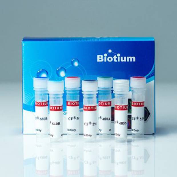
CF®543 Tyramide

Cat# 92172
Size : 500UG
Brand : Biotium
Contact local distributor :
 Phone : +1 850 650 7790
CF® Dye Tyramide
Fluorescent CF® Dye tyramides are used for tyramide signal amplification (TSA) for increasing immunofluorescence sensitivity in multicolor immunocytochemistry (ICC), immunohistochemistry (IHC), or in situ hybridization (ISH).
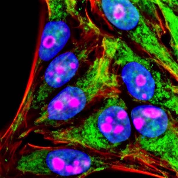 Sequential labeling of formaldehyde-fixed HeLa cells with two Tyramide Amplification Kits. Mitochondria (green) were imaged with rabbit anti-COXIV primary antibody and Tyramide Amplification Kit with HRP goat anti-rabbit IgG and CF®488A-tyramide, followed by peroxidase quenching. Nucleoli (magenta) were visualized with mouse anti-cyclin B1 primary antibody and Tyramide Amplification Kit with HRP Goat anti-mouse IgG and CF®568-tyramide. Actin (red) was detected with CF®640R-phalloidin, and cell nuclei (blue) were stained with DAPI.
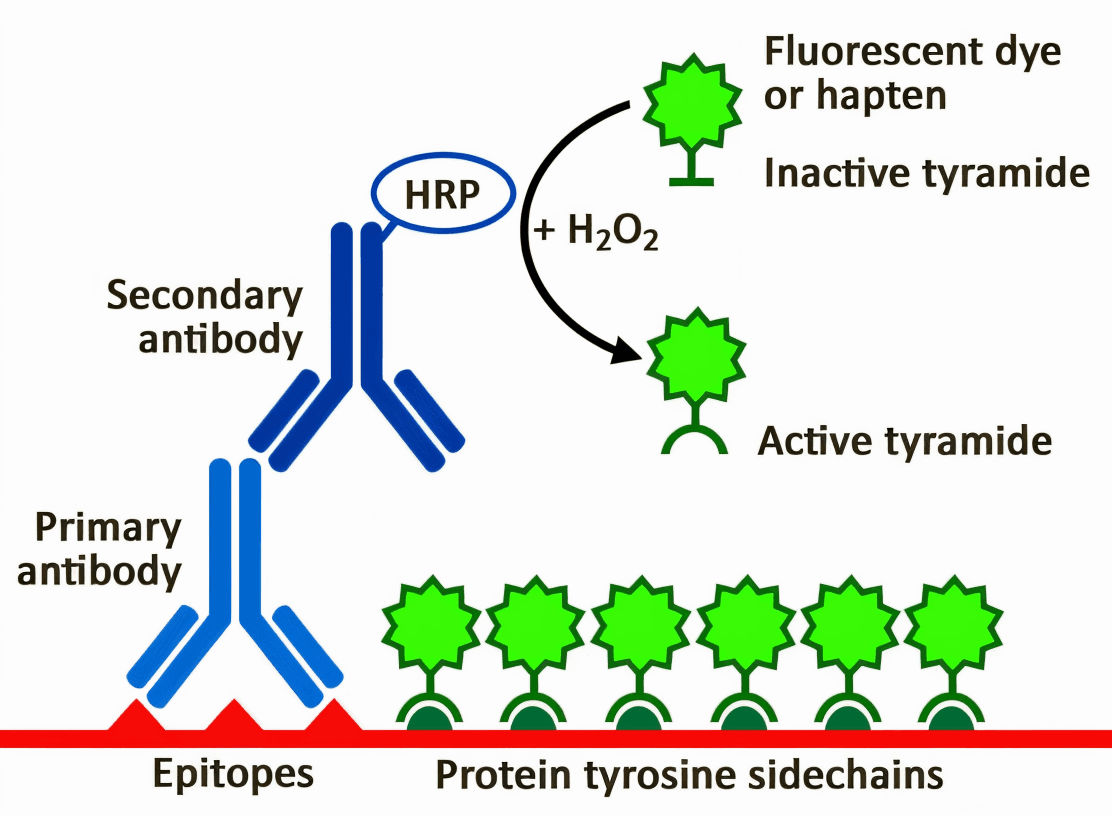 HRP-conjugated antibodies convert inactive tyramide to active tyramide, which then covalently attaches to nearby tyrosine residues. Source: Biotium
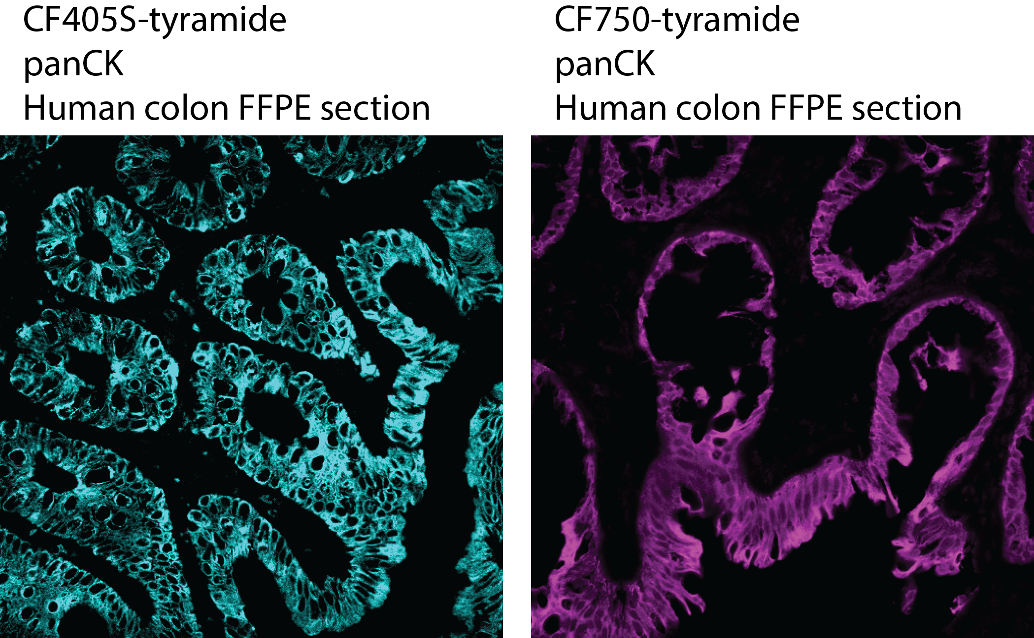 2uM of CF405S-tyramide (left) and 10uM of CF750-tyramide (right) staining human colon FFPE section labeled with mouse anti-panCK primary antibody and HRP-Goat anti-Mouse secondary antibody. The staining is performed using our Tyramide Enhancer Buffer.
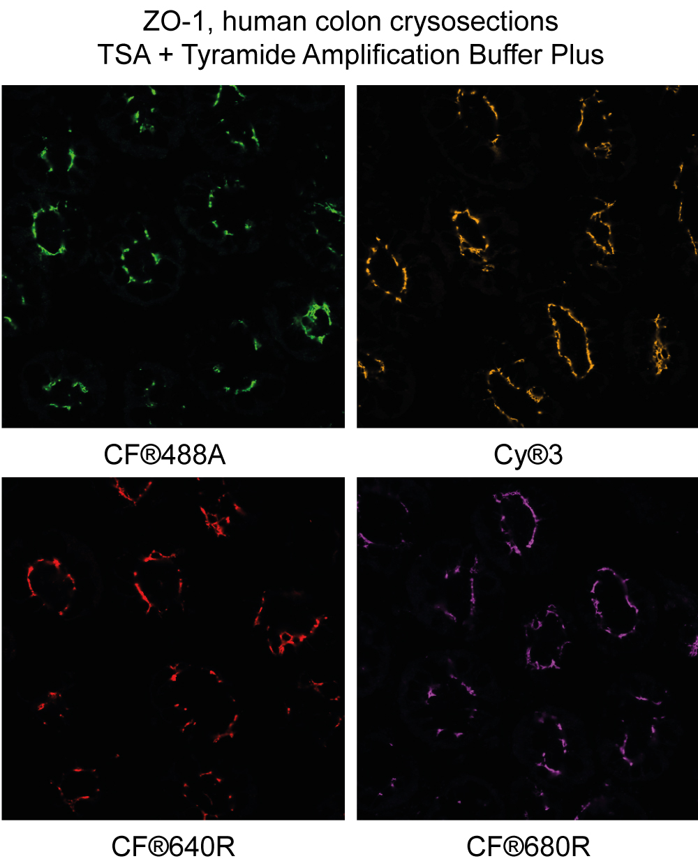 ZO-1 staining in human colon cryosections detected using CF®488A, Cyanine 555 (Cy®3), CF®640R, and CF®680R tyramide with Biotium's Tyramide Amplification Buffer Plus.
 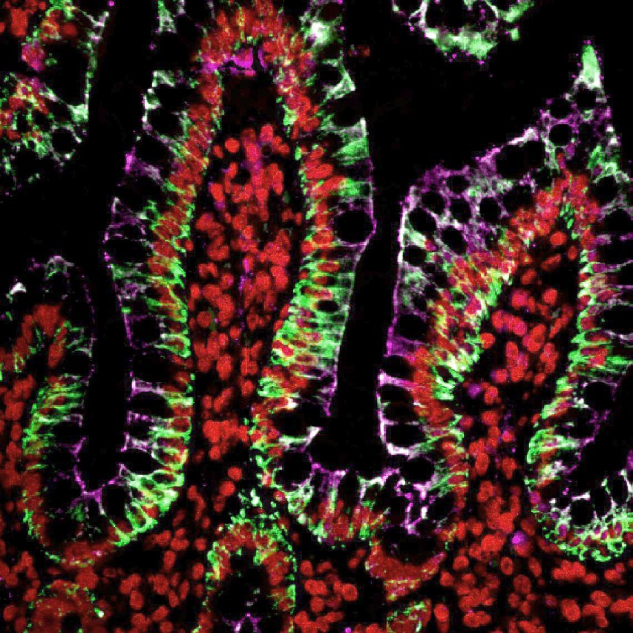 Multiplex tyramide labeling of FFPE tissues. Cytokeratin (pan) was labeled with CF®488A-tyramide (green); Histone H1 was labeled with Cy®3-tyramide (red); ZO1 was labeled with CF®640R-tyramide (magenta). Primary antibodies were from mouse, and secondary antibody was HRP-conjugated goat anti-mouse. Each labeling was performed sequentially, with antibody removal by microwave treatment between labeling steps.
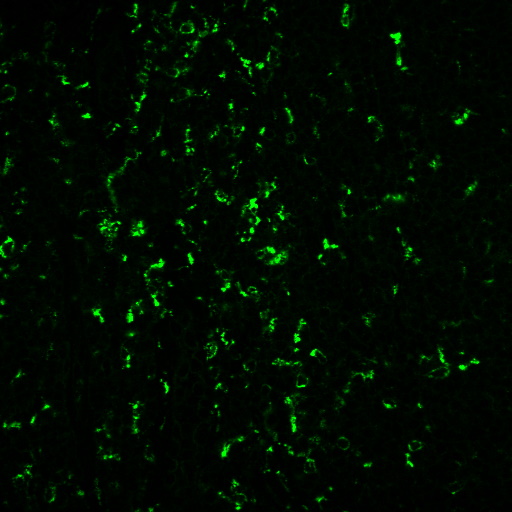 Human tonsil FFPE section stained with anti-CD68 (C68/684+KP1) and goat anti-mouse HRP, detected using CF®488A tyramide in Tyramide Amplification Buffer Plus.
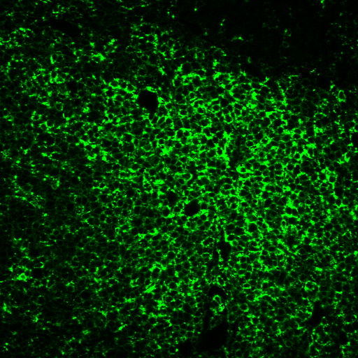 Human tonsil FFPE section stained with anti-CD45RO (UCHL-1) and goat anti-mouse HRP, detected using CF®488A tyramide in Tyramide Amplification Buffer Plus.
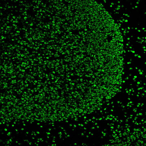 Human tonsil FFPE sections heated in AntiFix™ antigen retrieval buffer, then stained with anti-Ki67 MKI67/2466, goat anti-mouse HRP, and CF®488A tyramide using Tyramide Amplification Buffer Plus.
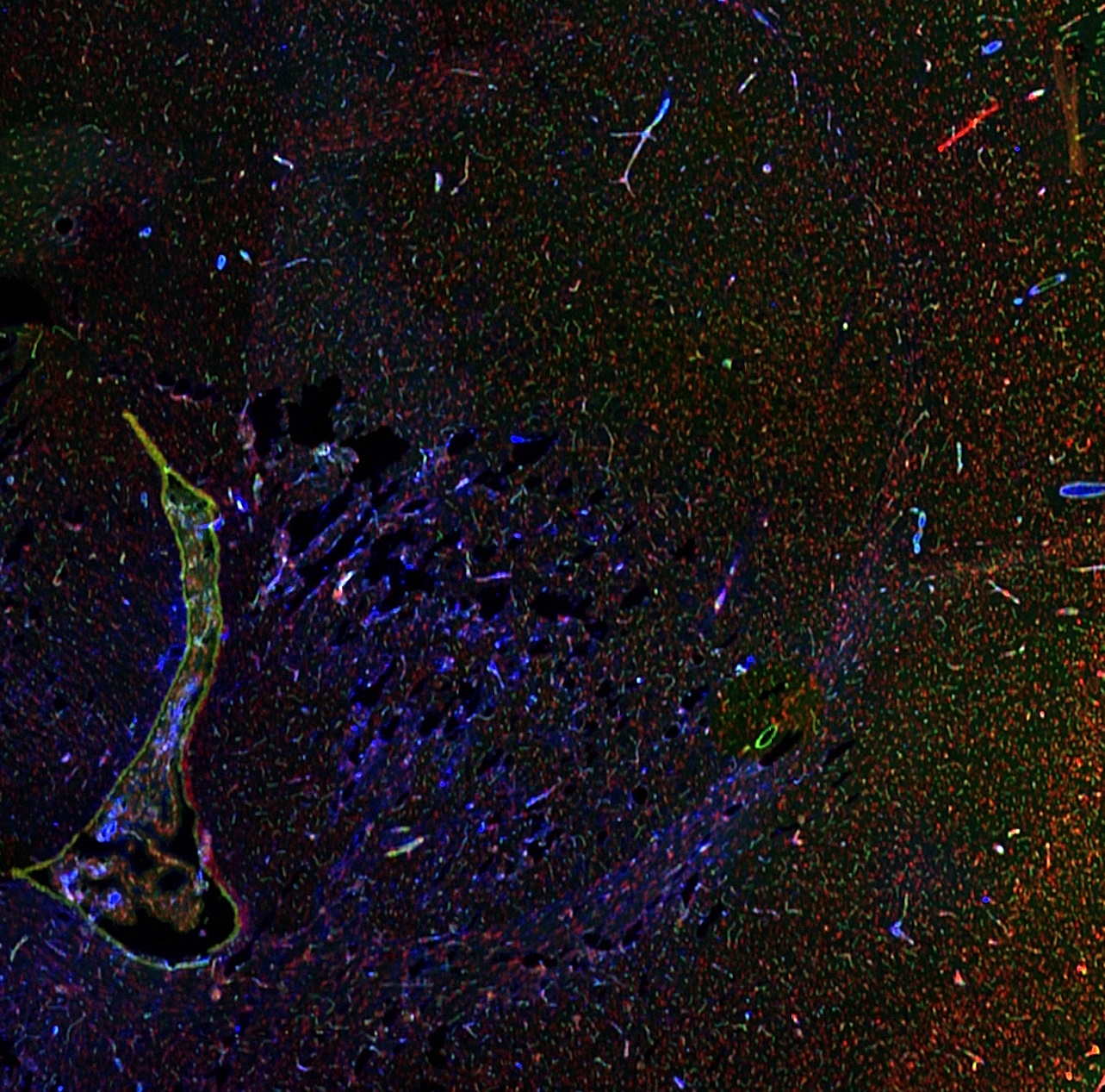 Section of rat brain and choroid plexus stained with mouse anti-GFAP GA5 antibody, HRP goat anti-mouse, and CF®740 Tyramide using Biotium's Tyramide Amplification Buffer Plus (glial cells, blue). Blood vessels were stained with CF®488A LEL Tomato Lectin (green), and nuclei were stained with NucSpot® 568/580 Nuclear Stain (red). Imaged on a LI-COR® Odyssey® M Imaging System in the 488, 520, and 800 channels.
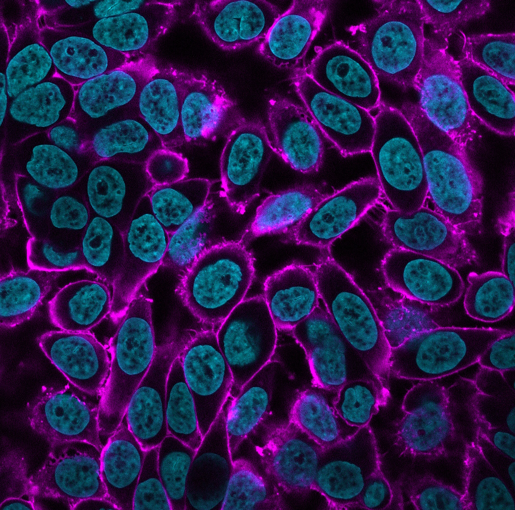 PFA-fixed HeLa cells stained with WGA-HRP detected with CF®740 Tyramide (magenta) in Biotium's Tyramide Amplification Buffer Plus. Nuclei were stained with Hoehcst (cyan). Imaged on an Evident FV4000 confocal imaging system with 730 nm laser line.
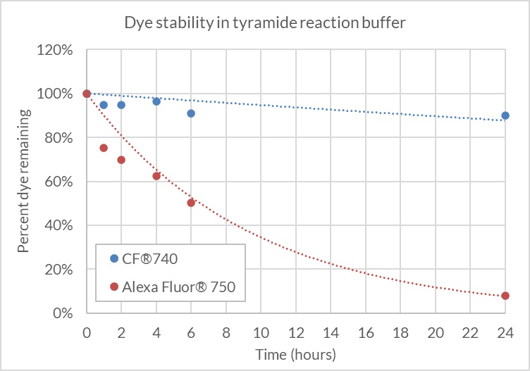 CF®740 free acid or Alexa Fluor® 750 free acid was diluted to 2 uM in 1X Tyramide Amplification Buffer with 0.0015% hydrogen peroxide and incubated at room temperature, protected from light. Dye concentration was measured by absorbance at the time points shown. CF®740 remained stable in the presence of hydrogen peroxide over 24 hours (≤90% of the starting dye concentration), while Alexa Fluor® 750 degraded to less than 10% of the starting concentration after 24 hours.
Product Description
CF® Dye tyramide conjugates are used for tyramide signal amplification (TSA), a method for high-density labeling of a target protein or nucleic acid in situ.
- High-density labeling of a target protein or nucleic acid for enhanced immunofluorescence sensitivity
- Especially suited for the detection of low abundance targets
- Detection sensitivity of over 100-fold compared to conventional procedures
- Enables multiplex multicolor detection, not limited by antibodies from the same host species
- Wide selection of bright, photostable and water-soluble CF® Dyes, excellent options for fluorescent labeling
- CF®740 Tyramide is a unique near-IR conjugate compatible with automated staining
Also learn about TyraMax™ Amplification Dyes and Kits, Biotium's next generation tyramide dyes that offer brighter signal compared to the original CF® Dye Tyramides, and have advantages in brightness, photostability, and working solution stability compared to other TSA dyes. We also offer Ready-to-Use Tyramide Amplification Buffer, Tyramide Amplification Buffer Plus (an improved formulation for enhanced TSA sensitivity), and CF® Dye Tyramide Amplification Kits.
Superior CF® Dyes
Biotium’s next-generation CF® Dyes were designed to be highly water-soluble with advantages in brightness and photostability compared to other commercially available fluorescent dyes. Our CF® Dye Tyramide conjugates are available in 22 colors. Learn more about CF® Dyes.
CF®740 Tyramide: A Stable Near-IR Tyramide for Automated Staining
For researchers considering near-infrared detection, we recommend CF®740 tyramide over CF®750 and CF®754. This is because CF®750 is unstable in oxidizing amplification buffer and should be added to the buffer immediately before performing the staining reaction. The poor stability in oxidizing amplification buffer makes the dye challenging to use for automated staining platforms (ie. BOND RX) that require longer periods with the dye in buffer. CF®754 tyramide is stable in oxidizing amplification buffer, but the dye has a broad absorption peak that can cause channel spillover. CF®740 tyramide was developed to be stable in oxidizing amplification buffer when compared to Alexa Fluor® 750 and CF®750. In addition, CF®740 tyramide has a narrower absorption peak that minimizes spillover and therefore a superior option to CF®754 tyramide. Near-IR tyramide background staining may be tissue dependent and not suitable for all targets. In general, we would recommend using near-IR staining for more abundant targets in your panel.
CF®740 free acid or Alexa Fluor® 750 free acid was diluted to 2 uM in 1X Tyramide Amplification Buffer with 0.0015% hydrogen peroxide and incubated at room temperature, protected from light. Dye concentration was measured by absorbance at the time points shown. CF®740 remained stable in the presence of hydrogen peroxide over 24 hours (≤90% of the starting dye concentration), while Alexa Fluor® 750 degraded to less than 10% of the starting concentration after 24 hours.
Tyramide Signal Amplification
TSA is a highly sensitive method for differential gene or protein analysis or detection of low-abundance targets, in fluorescent ICC, IHC, and FISH applications. An antibody- or streptavidin-HRP conjugate catalyzes the deposition of fluorescent dye/biotin tyramides on tyrosine residues on and adjacent to a target protein or nucleic acid sequence in situ. This results in high-density labeling of the target and significantly improves the detection sensitivity up to 100-fold compared to conventional methods. TSA is particularly advantageous for fluorescence detection in human tissue, where conventional ICC or FISH often fails to provide adequate signal over autofluorescence background. In applications where increased sensitivity is not required, TSA enables the use of significantly lower antibody or probe concentrations for the same level of detection sensitivity thereby reducing issues of non-specific binding or cross-reactivity. Furthermore, since binding of the tyramide label is covalent, a large number of targets can be detected in the same sample using multiple rounds of sequential TSA, in which the availability of antibodies from different host species is not a limitation. TSA also can be easily integrated with conventional immunostaining. Learn more about Tyramide Signal Amplification.
CF® Dye Tyramides
| Product | Ex/Em | MW (g/mol) | Size | Catalog No. | Dye Features |
|---|---|---|---|---|---|
| CF®350 Tyramide | 347/448 nm | ~614 | 0.5 mg | 92170 | CF®350 Features |
| CF®405S Tyramide | 404/431 nm | ~689 | 0.5 mg | 92197 | CF®405S Features |
| CF®405M Tyramide | 408/452 nm | ~621 | 0.5 mg | 96057 | CF®405M Features |
| CF®405L Tyramide | 395/545 nm | ~1692 | 0.5 mg | 92198 | CF®405L Features |
| CF®430 Tyramide | 426/498 nm | ~707 | 0.5 mg | 96053 | CF®430 Features |
| CF®488A Tyramide | 490/515 nm | ~666 | 0.5 mg | 92171 | CF®488A Features |
| CF®514 Tyramide | 516/548 nm | ~1337 | 0.5 mg | 92199 | CF®514 Features |
| CF®532 Tyramide | 527/558 nm | ~804 | 0.5 mg | 96066 | CF®532 Features |
| CF®543 Tyramide | 541/560 nm | ~1006 | 0.5 mg | 92172 | CF®543 Features |
| CF®550R Tyramide | 551/577 nm | ~806 | 0.5 mg | 96077 | CF®550R Features |
| CF®555 Tyramide | 555/565 nm | ~1120 | 0.5 mg | 96021 | CF®555 Features |
| CF®568 Tyramide | 562/583 nm | ~833 | 0.5 mg | 92173 | CF®568 Features |
| CF®583R Tyramide | 586/609 nm | ~892 | 0.5 mg | 96085 | CF®583R Features |
| CF®594 Tyramide | 593/614 nm | ~848 | 0.5 mg | 92174 | CF®594 Features |
| CF®620R Tyramide | 617/639 nm | ~857 | 0.5 mg | 92194 | CF®620R Features |
| CF®640R Tyramide | 642/662 nm | ~951 | 0.5 mg | 92175 | CF®640R Features |
| CF®647 Tyramide | 650/665 nm | ~1104 | 0.5 mg | 96022 | CF®647 Features |
| CF®660R Tyramide | 663/682 nm | ~1007 | 0.5 mg | 92195 | CF®660R Features |
| CF®680R Tyramide | 680/701 nm | ~1031 | 0.5 mg | 92196 | CF®680R Features |
| CF®710 Tyramide | 712/736 nm | ~977 | 0.5 mg | 96127 | CF®710 Features |
| CF®725 Tyramide | 729/750 nm | ~1005 | 0.5 mg | 96128 | CF®725 Features |
| CF®740 Tyramide | 742/767 nm | ~1005 | 0.5 mg | 96124 | CF®740 Features |
| CF®750 Tyramide* | 755/779 nm | ~3040 | 0.5 mg | 96052 | CF®750 Features |
| CF®754 Tyramide | 748/793 nm | ~1000 | 0.5 mg | 96090 |
* CF®750 Tyramide is not stable in TSA buffer, and should be added to the buffer immediately before performing the staining reaction.
Technology

Tech Tips

Product Attributes
Size
Chemical reactivity (reacts with)
Functional group
Assay type/options
Storage Conditions
Detection method/readout
Documents, Protocols, SDS and COA

Protocols
Datasheet
COA
Request the Certificate of AnalysisReferences
1. Journal of Immunology Research (2020), 2020:2328675. DOI:10.1155/2020/2328675
2. Scientific Reports (2019), 9:1144. DOI:10.1038/s41598-018-38171-5
3. bioRxiv (2019) preprint. DOI:10.1101/651083
4. OncoImmunology (2019), 8(6):e1581528-10. DOI:10.1080/2162402X.2019.1581528
5. Microbiological Research (2018), 217: 69-80. DOI:10.1016/j.micres.2018.08.017
6. Mol Neurodegeneration (2017), 12:68. DOI:10.1186/s13024-017-0202-z
7. Biology Open (2017), 6: 891-896. DOI:10.1242/bio.025809
8. The American Journal of Pathology (2016), 186(10):2650-2664. DOI:10.1016/j.ajpath.2016.06.020
Download a list of CF® Dye references.
Citations
1. Journal of Immunology Research (2020), 2020:2328675. DOI:10.1155/2020/2328675
2. Scientific Reports (2019), 9:1144. DOI:10.1038/s41598-018-38171-5
3. bioRxiv (2019) preprint. DOI:10.1101/651083
4. OncoImmunology (2019), 8(6):e1581528-10. DOI:10.1080/2162402X.2019.1581528
5. Microbiological Research (2018), 217: 69-80. DOI:10.1016/j.micres.2018.08.017
6. Mol Neurodegeneration (2017), 12:68. DOI:10.1186/s13024-017-0202-z
7. Biology Open (2017), 6: 891-896. DOI:10.1242/bio.025809
8. The American Journal of Pathology (2016), 186(10):2650-2664. DOI:10.1016/j.ajpath.2016.06.020
Download a list of CF® Dye references.
 Powered by Bioz