



The RayBiotech Immune Checkpoint Receptor Protein Array is a semi-quantitative array displaying 37 important human receptor proteins involved in immune checkpoint function.
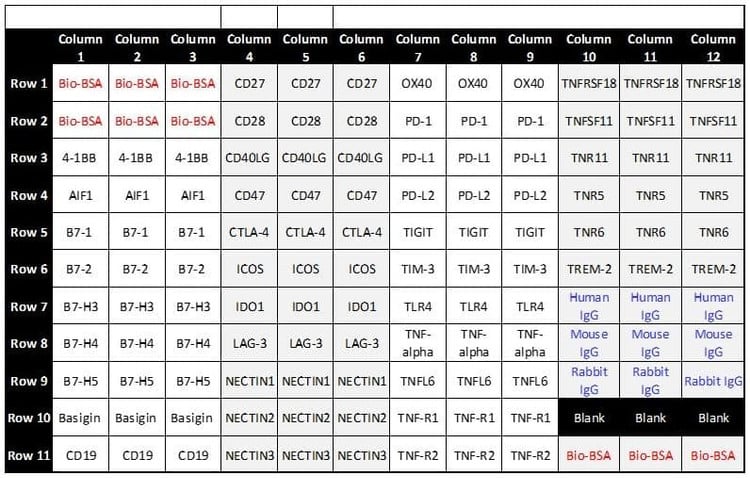
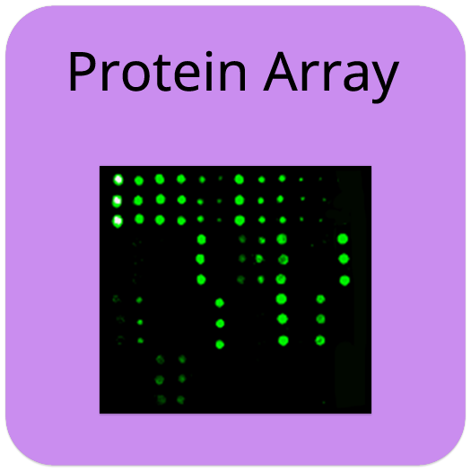
Human Immune Checkpoint Receptor Protein Array
Human Immune Checkpoint Receptor Protein Array (Human IgG)
Contact Us for Bulk Request
Product Description
Specifications
| Size | 1 slide (16 wells), 2 slides (32 wells) |
| Species | Human |
| Compatible Sample Types | Cell Culture Supernatants, Plasma, Serum, Ascites, Hybridoma cell culture medium |
| Quantitative/Semi-Quantitative | Semi-Quantitative |
| Number of Targets Detected | 37 |
| Solid Support | Glass Slide |
| Method Of Detection | Fluorescence Laser Scanner |
| Research Area | Cancer, Cancer Immunology, Adaptive Immunity |
| Shipping Type | Blue ice |
| Storage | -20°C |
| Antibody Detected | Human IgG, Rabbit IgG |
Introduction
The purified recombinant human receptor proteins are spotted in triplicate on a standard glass slide (25 mm x 75 mm x 1 mm), along with positive and negative controls to monitor each incubation step. When liquid sample types, such as serum or purified antibodies, are incubated on the array, "primary" antibodies in the sample will bind to their specific target receptor proteins. Specific primary antibody isotypes (IgG) are then targeted by biotin-conjugated anti-IgG secondary antibodies, respectively, that are specific for the host species. If the sample ligands/binders are not an antibody and the anti-ligand primary antibodies are available, the array test still can be conducted. If the sample ligands are biotin-conjugated, the array test will be more straight forward, easy and fast. See our product manual for details.
A fluorophore-conjugated streptavidin molecule is then added, which binds to the biotin on the secondary antibody. The fluorophore enables the detection of bound primary antibody via fluorescence using a laser scanner. The fluorescence signal is proportional to the amount of the immobilized receptor proteins. Since each spot represents a unique protein that is known, the specific target(s) bound by the ligands/antibodies can be ascertained.
Target Names
Totally 37 essential human receptor proteins playing the critical roles in immune checkpoints were selected, see below. All proteins are recombinant, expressed in mammalian cells with abundant post-translation modifications (PTMs), and affinity purified with high purity. The expressed regions are either the full-length proteins or the extracellular cellular membrane domains (ECD).
| Protein | Gene Symbol | UniProt Accession # |
|---|---|---|
| 4-1BB, TNFRSF9 | TNFRSF9, CD137, ILA | Q07011 |
| AIF1 | AIF1, G1, IBA1 | P55008 |
| B7-1 | CD80, CD28LG, CD28LG1, LAB7 | P33681 |
| B7-2 | CD86, CD28LG2 | P42081 |
| B7-H3 | CD276, B7H3, PSEC0249, UNQ309/PRO352 | Q5ZPR3 |
| B7-H4, VTCN1 | VTCN1, B7H4, UNQ659/PRO1291 | Q7Z7D3 |
| B7-H5, VISTA | VSIR, C10orf54, SISP1, VISTA, PP2135, UNQ730/PRO1412 | Q9H7M9 |
| Basigin | BSG, UNQ6505/PRO21383 | P35613 |
| CD19 | CD19 | P15391 |
| CD27/TNFRSF7 | CD27, TNFRSF7 | P26842 |
| CD28 | CD28 | P10747 |
| CD40LG, CD40L | CD40LG, CD40L, TNFSF5, TRAP | P29965 |
| CD47 | CD47, MER6 | Q08722 |
| CTLA-4 | CTLA4, CD152 | P16410 |
| ICOS | ICOS, AILIM | Q9Y6W8 |
| IDO1 | IDO1, IDO, INDO | P14902 |
| LAG-3 | LAG3, FDC | P18627 |
| NECTIN1 | NECTIN1, HVEC, PRR1, PVRL1 | Q15223 |
| NECTIN2 | NECTIN2, HVEB, PRR2, PVRL2 | Q92692 |
| NECTIN3 | NECTIN3, PRR3, PVRL3 | Q9NQS3 |
| OX40, TNFRSF4 | TNFRSF4, TXGP1L | P43489 |
| PD-1 | PDCD1, PD1 | Q15116 |
| PD-L1 | CD274, B7H1, PDCD1L1, PDCD1LG1, PDL1 | Q9NZQ7 |
| PD-L2 | PDCD1LG2, B7DC, CD273, PDCD1L2, PDL2 | Q9BQ51 |
| TIGIT | TIGIT, VSIG9, VSTM3 | Q495A1 |
| TIM-3 | HAVCR2, TIM3, TIMD3 | Q8TDQ0 |
| TLR4 | TLR4 | O00206 |
| TNF-alpha | TNF, TNFA, TNFSF2 | P01375 |
| TNFL6 | FASLG, APT1LG1, CD95L, FASL, TNFSF6 | P48023 |
| TNF-R1 | TNFRSF1A, TNFAR, TNFR1 | P19438 |
| TNF-R2 | TNFRSF1B, TNFBR, TNFR2 | P20333 |
| TNFRSF18 | TNFRSF18, AITR, GITR, UNQ319/PRO364 | Q9Y5U5 |
| TNFSF11, OPGL, RANKL | TNFSF11, OPGL, RANKL, TRANCE | O14788 |
| TNR11, TNFRSF11 | TNFRSF11A, RANK | Q9Y6Q6 |
| TNR5, TNFRSF5 | CD40, TNFRSF5 | P25942 |
| TNR6, TNFRSF6 | FAS, APT1, FAS1, TNFRSF6 | P25445 |
| TREM-2 | TREM2 | Q9NZC2 |
Application Notes
- Detection of antibody binding to different immune checkpoint proteins from serum or plasma samples
- Therapeutical drug discovery and development: Screening and characterization of small binder molecules, types of antibodies (polyclonal and monoclonal antibodies, scFv, VHH, etc.), or biologics that target immune checkpoints.
- Biomarker discovery: Identifying new biomarkers for immune checkpoint activity or dysfunction.
- Mechanistic studies: Understanding signaling pathways and receptor interactions in the immune system.
- Clinical research: Evaluating patient-specific responses to immune checkpoint inhibitors in cancer therapy.
Product Features
- Small sample volume required
- High density
- High sensitivity
- Large dynamic range
- Suitable for high-throughput assays
Kit Components
- RayBio® Human Receptor Protein Glass Array
- Blocking Buffer
- Wash Buffer I
- Wash Buffer II
- Biotin-conjugated secondary antibody
- Cy3 equivalent dye conjugated streptavidin
- Array accessories (including slide incubation chamber assembly, gasket, adhesive plastic strips, etc.)
Other Materials Required
- Distilled water
- Small plastic boxes or containers
- Orbital shaker or oscillating rocker
- Pipettors, pipette tips and other common lab consumables
- Aluminum foil
- Laser fluorescence scanner
View Compatible Laser Scanners
Don't have a compatible scanner? RayBiotech now offers FREE scanning service for all RayBio glass slide antibody arrays! Learn More
Storage/Stability
Upon receipt, all kit components should be stored at -20°C to -80°C until ready to use. Stored properly, the kit will retain complete activity for up to 6 months.
documents
GAL (GAL-PAH-ICP-G1.gal)
Excel (PAH-ICP-G1_Analysis_Tool.xlsm)