



Detects 18 different phosphorylated human and mouse proteins crucial to the AKT Pathway

Human/Mouse AKT Pathway Phosphorylation Array C1
Human/Mouse AKT Pathway Phosphorylation Array C1
Contact Us for Bulk Request
Product Description
Specifications
| Size | 2 Sample Kit, 4 Sample Kit, 8 Sample Kit |
| Species | Human, Mouse |
| Compatible Sample Types | Cell Culture Supernatants, Plasma, Serum, Tissue Lysates, Cell Lysates |
| Quantitative/Semi-Quantitative | Semi-Quantitative |
| Number of Targets Detected | 18 |
| Recommended Applications | Multiplexed Protein Detection; Detection of Relative Protein Expression; Detecting Patterns of Cytokine Expression; Biomarker/ Key Factor Screening; Identifying Key Factors; Confirming a Biological Process |
| Solid Support | Membrane |
| Design Principle | Sandwich-based |
| Method Of Detection | Chemiluminescence |
| Research Area | Post-Translational Modifications, Phosphorylation, AMPK Signaling, HER/ErbB Signaling, HIF-1 alpha Signaling, IGF Signaling, JAK/STAT Signaling, MAPK Signaling, mTOR Signaling, PI3K-AKT Signaling, TGF-beta Signaling, Wnt-beta-Catenin Signaling, Akt Signaling |
| Shipping Type | Blue ice |
| Storage | -20°C |
Related Products
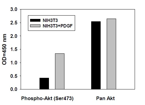
Phospho-AKT (S473) ELISA Kit
As low as $521.00
Product Features
- Easy to use
- No specialized equipment needed
- Compatible with nearly any liquid sample
- Proven technology (many publications)
- Highly sensitive
- Sandwich ELISA specificity
- Higher density than ELISA, Western blot or bead-based multiplex
Target Names
| Scroll over each target protein for more information | ||||
|---|---|---|---|---|
| Akt (P-Ser473) (PKB (P-Ser473))
| AMPKa (P-Thr172)
| BAD (P-Ser112)
| 4E-BP1 (P-Thr36)
| ERK1 (P-T202/Y204)/ERK2 (P-Y185/Y187)
|
| GSK3a (P-Ser21)
| GSK3b (P-Ser9)
| mTOR (P-Ser2448)
| p27 (P-Thr198)
| P53 (P-Ser15)
|
| P70S6K (P-Thr421/Ser424)
| PDK1 (P-Ser241)
| PRAS40 (P-Thr246)
| PTEN (P-Ser380)
| Raf-1 (Ser301)
|
| RPS6 (P-Ser235/Ser236)
| RSK1 (P-Ser380)
| RSK2 (P-Ser386)
| ||
Kit Components
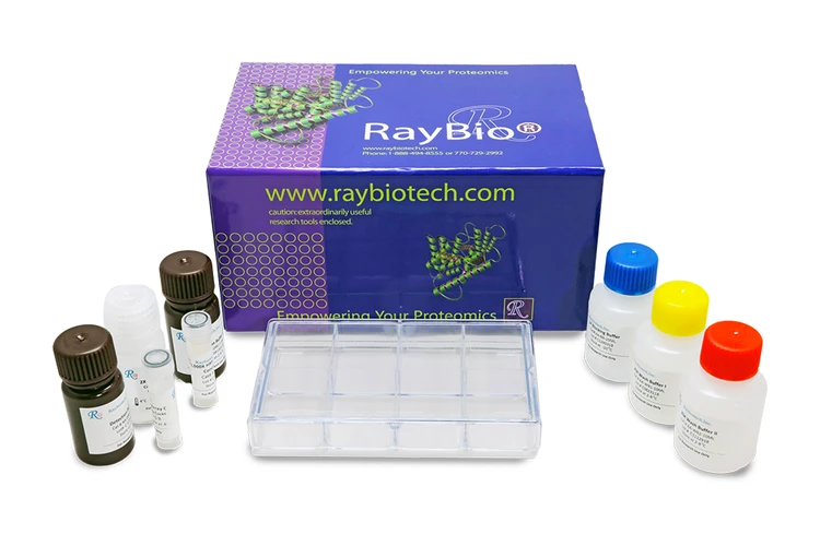
- Human and Mouse AKT Pathway Phosphorylation Antibody Array C1 Membranes
- Blocking Buffer
- Detection Antibody Cocktail
- 1,000X HRP-Anti-Rabbit IgG Concentrate
- 20X Wash Buffer I Concentrate
- 20X Wash Buffer II Concentate
- 2X Cell Lysis Buffer Concentrate
- Detection Buffer C
- Detection Buffer D
- 8-Well Incubation Tray
- Protease Inhibitor Cocktail
- 100x Phosphatase Inhibitor Cocktail I
- Phosphatase Inhibitor Cocktail II
- Plastic Sheets
- Array Map Template
- Manual
Additional Materials Required
- Pipettors, pipet tips and other common lab consumables
- Orbital shaker or oscillating rocker
- Tissue Paper, blotting paper or chromatography paper
- Adhesive tape or Saran Wrap
- Distilled or de-ionized water
- A chemiluminescent blot documentation system (such as UVP's ChemiDoc-It® or EpiChem II Benchtop Darkroom or GE's ImageQuant™ LAS 4000 or Amersham Imagers 600 and 680), X-ray Film and a suitable film processor, or another chemiluminescent detection system.
Assay Procedure Summary
- Block membranes
- Incubate with Sample
- Incubate with Detection Antibody Cocktail
- Incubate with HRP-Conjugated anti-IgG
- Incubate with Detection Buffers
- Image with chemiluminescent imaging system
- Perform densitometry and analysis
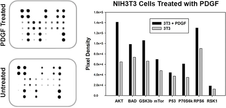
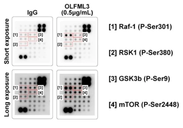
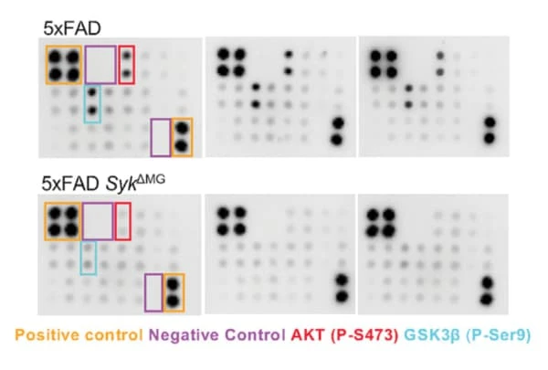
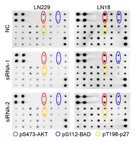
Typical Data

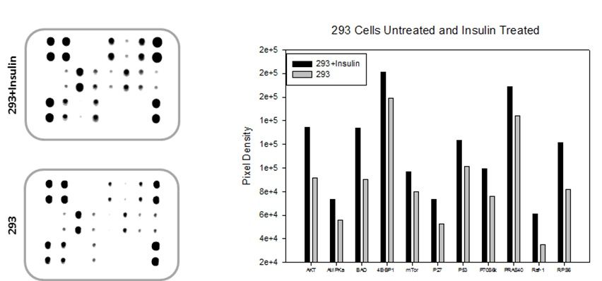
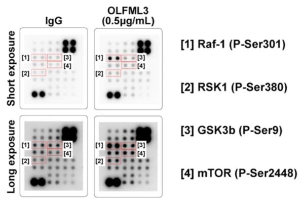
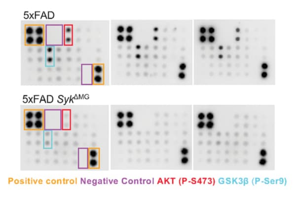
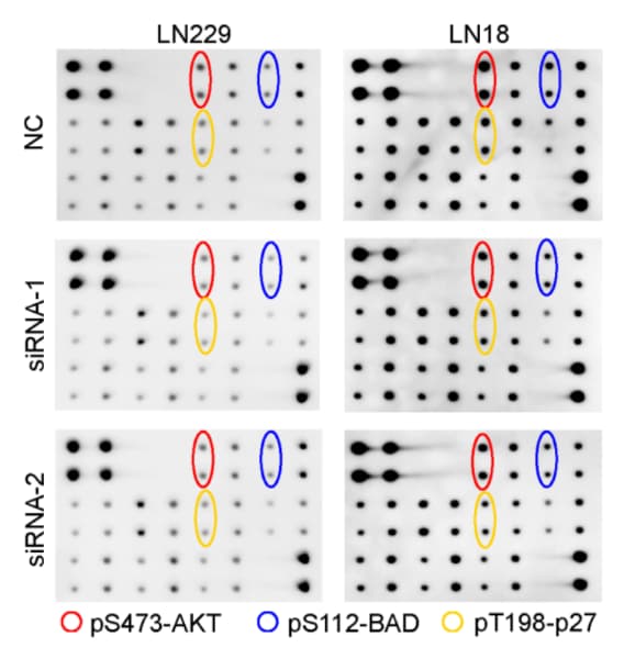
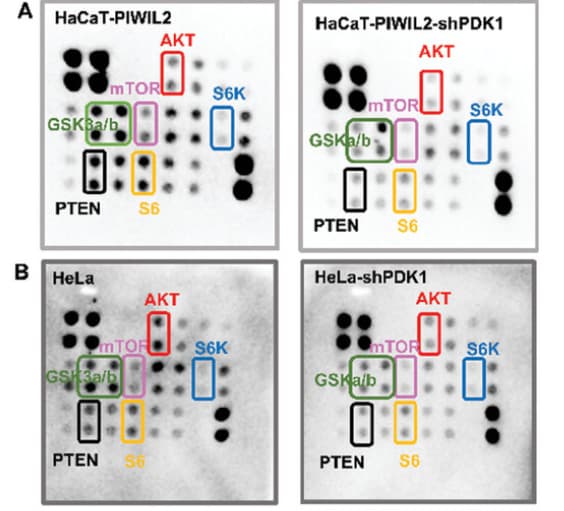
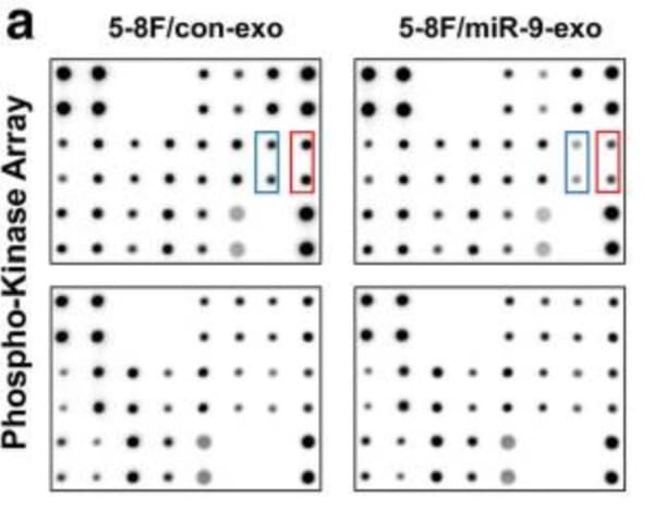
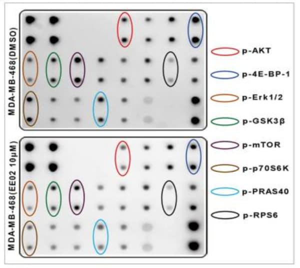
Storage/Stability
For best results, store the entire kit frozen at -20°C upon arrival. Stored frozen, the kit will be stable for at least 6 months which is the duration of the product warranty period. Once thawed, store array membranes and 1X Blocking Buffer at -20°C and all other reagents undiluted at 4°C for no more than 3 months.
documents
citations
SYK coordinates neuroprotective microglial responses in neurodegenerative disease
- Year:
- 2022
- Journal Name:
- Cell
- PubMed ID:
- 36257314
SVEP1 is an endogenous ligand for the orphan receptor PEAR1
- Species:
- Human
- Sample Type:
- human umbilical vein endothelial cells (HUVECs)
- Year:
- 2023
- Journal Name:
- Nature Communications
NLRP6 potentiates PI3K/AKT signalling by promoting autophagic degradation of p85α to drive tumorigenesis
- Species:
- Human
- Sample Type:
- HEK293T cell
- Year:
- 2023
- Journal Name:
- Nature Communications
LIN28B-mediated PI3K/AKT pathway activation promotes metastasis in colorectal cancer models
- Year:
- 2025
- Journal Name:
- Journal of Clinical Investigation
- PubMed ID:
- 39808497
Activated mTOR Signaling in the RPE Drives EMT, Autophagy, and Metabolic Disruption, Resulting in AMD-Like Pathology in Mice
- Year:
- 2025
- Journal Name:
- Aging Cell
- PubMed ID:
- 39957408
Epithelial Genetic Muscarinic Receptor 3 Ablation Induces Sex-Specific Modulation of Colonic Intestinal Progenitor Cells and Response to Intestinal Injury
- Year:
- 2025
- Journal Name:
- Journal of Crohns & Colitis
- PubMed ID:
- 40052554
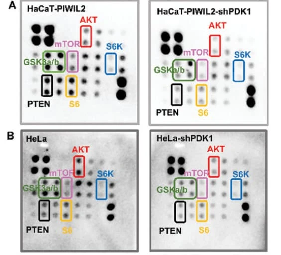
PIWIL2/PDK1 Axis Promotes the Progression of Cervical Epithelial Lesions via Metabolic Reprogramming to Maintain Tumor-Initiating Cell Stemness
- Year:
- 2024
- Journal Name:
- Advanced science (Weinheim, Baden-Württemberg, Germany)
- PubMed ID:
- 39499767
LAMA5-inspired adhesive dodecapeptide facilitates efficient dentine regeneration: An in vitro and in vivo study
- Species:
- Human
- Sample Type:
- human dental pulp cells (hDPCs)
- Year:
- 2023
- Journal Name:
- International Endodontic Journal
Exosome-transmitted circCABIN1 promotes temozolomide resistance in glioblastoma via sustaining ErbB downstream signaling
- Species:
- Human
- Sample Type:
- LN229
- Year:
- 2023
- Journal Name:
- Journal of nanobiotechnology
YBX1/lncRNA SBF2-AS1 interaction regulates proliferation and tamoxifen sensitivity via PI3K/AKT/MTOR signaling in breast cancer cells
- Species:
- Human
- Sample Type:
- MCF-7 cell
- Year:
- 2023
- Journal Name:
- Molecular Biology Reports
