



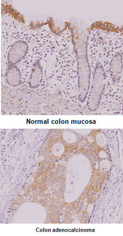
Immunohistochemistry (Formalin/PFA-fixed paraffin-embedded sections)
Immunohistochemistry analysis of normal colon mucosa and colon adenocalcinoma tissue were incubated with IRF5 monoclonal antibody, clone 10T1 (Cat# MAB1068) (1:50) for 2 hours at room temperature, was performed in 0.1M sodium citrate buffer and detected using Diaminobenzidine (DAB).
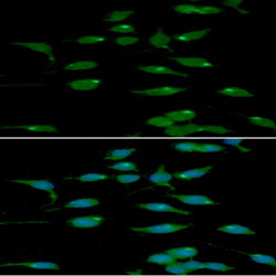
Immunofluorescence
Immunofluorescence analysis of Raw264.7 cells. The cell was stained with IRF5 monoclonal antibody, clone 10T1 (Cat# MAB1068) (1:100). The secondary antibody (green) was used Alexa Fluor 488. DAPI was stained the cell nucleus (blue).
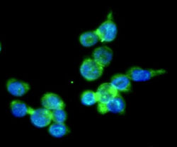
Immunocytochemistry
Immunocytochemistry analysis of THP-1 cells. The cell was stained with IRF5 monoclonal antibody, clone 10T1 (Cat#MAB1068) (1:100). The secondary antibody (green) was used Alexa Fluor 488. DAPI was stained the cell nucleus (blue).
- Specifications
Product Description
Mouse monoclonal antibody raised against partial recombinant IRF5.
Immunogen
Recombinant protein corresponding to amino acids 176-240 of human IRF5.
Host
Mouse
Reactivity
Human, Mouse
Form
Liquid
Purification
Protein A purification
Isotype
IgG1, kappa
Recommend Usage
ELISA
Flow Cytometry
Immunocytochemistry
Immunofluorescence
Immunohistochemistry
Western Blot
The optimal working dilution should be determined by the end user.Storage Buffer
In PBS, pH 7.4 (10% glycerol, 0.02% sodium azide).
Storage Instruction
Store at 2°C to 8°C for 1 week. For long term storage, aliquot and store at -20°C to -80°C.
Aliquot to avoid repeated freezing and thawing. - Applications
Immunohistochemistry (Formalin/PFA-fixed paraffin-embedded sections)
Immunohistochemistry analysis of normal colon mucosa and colon adenocalcinoma tissue were incubated with IRF5 monoclonal antibody, clone 10T1 (Cat# MAB1068) (1:50) for 2 hours at room temperature, was performed in 0.1M sodium citrate buffer and detected using Diaminobenzidine (DAB).Immunofluorescence
Immunofluorescence analysis of Raw264.7 cells. The cell was stained with IRF5 monoclonal antibody, clone 10T1 (Cat# MAB1068) (1:100). The secondary antibody (green) was used Alexa Fluor 488. DAPI was stained the cell nucleus (blue).Immunocytochemistry
Immunocytochemistry analysis of THP-1 cells. The cell was stained with IRF5 monoclonal antibody, clone 10T1 (Cat#MAB1068) (1:100). The secondary antibody (green) was used Alexa Fluor 488. DAPI was stained the cell nucleus (blue).Flow Cytometry
Flow cytometry analysis of THP-1 cells, staining at 2-5ug for 1x10⁶cells (red line). The secondary antibody used goat anti-mouse IgG Alexa fluor 488 conjugate. Isotype control antibody was mouse IgG (black line).Enzyme-linked Immunoabsorbent Assay
Western Blot (Cell lysate)
Western blot analysis of Lane 1: Ramos cell lysate; Land 2: THP-1 cell lysate; Lane 3: A20 cell lysate; Lane 4: NIH3T3 cell lysate. - Gene Info — IRF5
Entrez GeneID
3663GeneBank Accession#
NM_002200Protein Accession#
NP_002191Gene Name
IRF5
Gene Alias
SLEB10
Gene Description
interferon regulatory factor 5
Omim ID
607218Gene Ontology
HyperlinkGene Summary
This gene encodes a member of the interferon regulatory factor (IRF) family, a group of transcription factors with diverse roles, including virus-mediated activation of interferon, and modulation of cell growth, differentiation, apoptosis, and immune system activity. Members of the IRF family are characterized by a conserved N-terminal DNA-binding domain containing tryptophan (W) repeats. Alternative splice variants encoding different isoforms exist. [provided by RefSeq
Other Designations
-
- Interactomes
- Pathways
- Diseases
- Publication Reference
- Rotavirus NSP1 inhibits expression of type I interferon by antagonizing the function of interferon regulatory factors IRF3, IRF5, and IRF7.
Barro M, Patton JT.
Journal of Virology 2007 May; 81(9):4473.
Application:WB-Tr, Human, HEK 293T cells.
- Interferon regulatory factor 5 represses expression of the Epstein-Barr virus oncoprotein LMP1: braking of the IRF7/LMP1 regulatory circuit.
Ning S, Huye LE, Pagano JS.
Journal of Virology 2005 Sep; 79(18):11671.
- Rotavirus NSP1 inhibits expression of type I interferon by antagonizing the function of interferon regulatory factors IRF3, IRF5, and IRF7.