
Mouse CD24/HSA Antibody : LE/AF

Cat# OASB00632
Size : 0.5mg
Brand : Aviva Systems Biology
Contact local distributor :

Phone : +1 850 650 7790
Mouse CD24/HSA Antibody : LE/AF (OASB00632)
| Datasheets/Manuals | Printable datasheet for Mouse CD24/HSA Antibody : LE/AF (OASB00632) |
|---|
| Tested Species Reactivity | Mouse |
|---|---|
| Predicted Species Reactivity | Mouse |
| Clonality | Monoclonal |
| Clone | 30-F1 |
| Isotype | IgG2c |
| Host | Rat |
| Conjugation | LE: Low Endotoxin, AF: Azide Free |
| Application | FC |
| Additional Information | Description: CD24 is expressed in B cells until their terminal differentiation into plasma cells. It has been used widely as a marker to fractionate different B-lineage differentiation stages. CD24 has been found on a variety of other hematopoietic and neural cell types and is now classified as a costimulatory molecule. The CD24 molecule, also known as HSA, is composed mostly of carbohydrate with a protein core of only 31-35 amino acids in the human and 27 amino acids in the mouse. CD24 is now included in the large group of phosphatidylinositol-anchored membrane proteins. Studies with purified CD24 applied to latex beads have demonstrated a self-binding property suggesting that the molecule is capable of promoting homotypic aggregation, binding, and cell adhesion. This is supported by earlier observations of the blockade of B cell lymphoblast aggregation with an anti-CD24 monoclonal antibody. Recent studies of murine CD24 have assigned a role for CD24 as a ligand for P-selectin, a calcium-dependent lectin. |
| Reconstitution and Storage | Store at -20C |
| Immunogen | Mouse thymus or spleen |
| Concentration | 0.5 mg/mL |
| Specificity | CD24 |
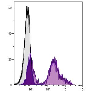
| Characterization | To ensure lot- to- lot consistency, each batch of monoclonal antibody is tested by flow cytometry to conformto characteristics of a standard reference reagent. Representative data are included in this product insert. |
| Warning | Reagents contain sodium azide. Sodium azide is very toxic if ingested or inhaled. Avoid contact withskin, eyes, or clothing. Wear eye or face protection when handling. If skin or eye contact occurs, washwith copious amounts of water. If ingested or inhaled, contact a physician immediately. Sodium azideyields toxic hydrazoic acid under acidic conditions. Dilute azide- containing compounds in running waterbefore discarding to avoid accumulation of potentially explosive deposits in lead or copper plumbing. |
| Dilution | Flow Cytometry: Fluorescein conjugate <= 2 ug/106 cells Biotin conjugate <= 2 ug/106 cells R-PE and APC conjugates <= 0.2 ug/106 cells Spectral Red conjugate <= 0.2 ug/106 cells |
| Application Info | Flow cytometry and cell sorting 8,10 |
| Other Applications Data | Since applications vary, you should determine the optimum working dilution for the product that is appropriate for yourspecific need. |
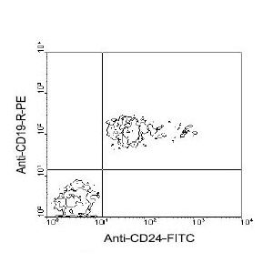
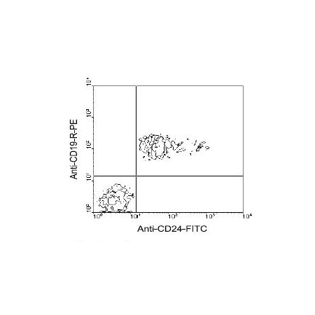
| Other Applications Image 1 Data | Amount Used: 3 Ug/106 cells BALB/c spleen cells were double-stained with rat anti-mouse CD24-FITC and rat anti-mouse CD19-RPE , following which small lymphocytes were gated and analyzed on a FACScan flow cytometer (BDIS, San Jose, CA). |
| Storage | - The purified (UNLB) antibody is supplied as 0.5 mg ofpurified immunoglobulin in 1.0 mL of 100 mM borate buffered saline, pH 8.2. No preservatives oramine- containing buffer salts added. Store at 2- 8 C - The fluorescein (FITC) conjugate is supplied as 0.5 mg or 0.1 mg in 1.0 mL of PBS/NaN3. Store at 2- 8 C - The biotin (BIOT) conjugate is supplied as 0.5 mg in 1.0 mL of PBS/NaN3. Store at 2- 8 C - The R- phycoerythrin (R- PE) and allophycocyanin (APC) conjugates are supplied as 0.1 mg in 1.0 mLor 0.2 mg in 2.0 mL of PBS/NaN3 and a stabilizing agent. Store at 2- 8 C. Do not freeze! - The Spectral Red (SPRD) conjugate is supplied as 0.1 mg in 1.0 mL of PBS/NaN3 and a stabilizingagent. Store at 2- 8 C. Do not freeze! - The low endotoxin, azide- free (LE/AF) antibody is supplied as 0.5 mg purified immunoglobulin in 1.0 mLof PBS. Aliquot and store at or below - 20 C - Protect conjugated forms from light. Aliquot and freeze the low endotoxin, azide- free product at - 20 Cimmediately upon receipt. Each reagent is stable for the period shown on the bottle label if stored asdirected. |
| Reference | 1. Kay, R., P. M. Rosten and R. K. Humphries. 1991. J. Immunol. 147:1412- 1416 2. Fischer, G. F., O. Majdic, S. Gadd, and W. Knapp. 1990. J. Immunol. 144:638 3. Hubbe, M. and P. Altevogt. 1994. Eur. J. Immunol. 1994. 24:731 4. Liu, Y., B. Jones, W. Brady, J. C. A. Janeway, and P. S. Linsley. 1992. Eur. J. Immunol. 22:2855 5. Shields, J. G., S. H. Smith and R. E. Callard. 1987. In Leukocyte Typing III. A. J. McMichael, et. al., eds., Oxford University Press, Oxford. P. 488 6. Kadmon, G., M. Eckert, M. Sammar, M. Schachner, and P. Altevogt. 1992. J. Cell Biol. 118:1245 7. Aigner, S., M. Ruppert, M. Hubbe, M. Sammar, Z. Sthoeger, E. C. Butcher, D. Vestweber, and P. Altevogt. 1995. International Immunology 7:1557 8. Ledbetter, J. A., and L. A. Herzenberg. 1979. Immunol. Rev. 47: 63 9. Kariv, I., R. R. Hardy and K. Hayakawa. 1994. J. Exp. Med. 180:2419 10. Hardy, R. R., C. E. Carmack, S. A. Shinton, J. D. Kemp, and K. Hayakawa. 1991. J. Exp. Med 173:1213. |
|---|---|
| Gene Symbol | CD24A |
| Gene Full Name | Signal transducer CD24 |
| Alias Symbols | H, HSA, Cd24, Ly-5, nect, Ly-52, nectadrin |
| NCBI Gene Id | 12484 |
| Protein Name | signal transducer CD24 |
| Uniprot ID | P24807 |
| Protein Accession # | NP_033976.1 |
| Nucleotide Accession # | NM_009846.2 |