

Orcein [1400-62-0]

Cat# T19835-5mg
Size : 5mg
Brand : TargetMol
Contact local distributor :
 Phone : +1 850 650 7790
Orcein
Copy Product InfoOrcein is a natural dye derived from lichen species, composed of a mixture of phenazone compounds that bind to chromatin through electrostatic and hydrophobic interactions at acidic pH. For fluorescence microscopy, orcein solutions exhibit emission peaks at 587 nm at low concentrations and 620 nm at high concentrations, with stained chromatin showing an emission peak at 585 nm and a shoulder at 620 nm. In light microscopy, acetic orcein remains a standard method for visualizing polytene chromosome banding in Drosophila salivary glands, enabling high-resolution analysis of interphase chromatin structure after endoreplication. Additionally, orcein selectively stains elastic fibers and has been used to evaluate fibrosis quality in liver tissue, where its staining pattern helps distinguish progressive from regressive fibrosis.
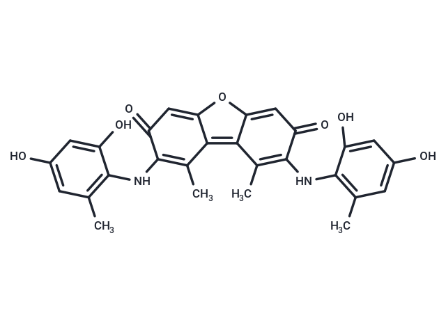
Cas No. 1400-62-0
For research use only—not for human use. No sales to individuals. Use as intended only.
Product Information
Bioactivity
Chemical Properties
Storage & Solubility Information
| Description | Orcein is a natural dye derived from lichen species, composed of a mixture of phenazone compounds that bind to chromatin through electrostatic and hydrophobic interactions at acidic pH. For fluorescence microscopy, orcein solutions exhibit emission peaks at 587 nm at low concentrations and 620 nm at high concentrations, with stained chromatin showing an emission peak at 585 nm and a shoulder at 620 nm. In light microscopy, acetic orcein remains a standard method for visualizing polytene chromosome banding in Drosophila salivary glands, enabling high-resolution analysis of interphase chromatin structure after endoreplication. Additionally, orcein selectively stains elastic fibers and has been used to evaluate fibrosis quality in liver tissue, where its staining pattern helps distinguish progressive from regressive fibrosis. |
| In vivo | Orcein is used in mouse models for histochemical staining of elastic fibers in atherosclerotic plaques. In apoE/LDLR-/- mice, orcein combined with martius scarlet blue (OMSB) staining enables qualitative and quantitative analyses of brachiocephalic artery sections, identifying elastic fibers (purple), collagen (blue), erythrocytes (yellow), and fibrin (red). The method is simple, under 1 hour, and compatible with automated stainers for digital segmentation. |
| Molecular Weight | 500.507 |
| Formula | C28H24N2O7 |
| Cas No. | 1400-62-0 |
| Smiles | CC1=C(Nc2c(C)cc(O)cc2O)C(=O)C=c2oc3=CC(=O)C(Nc4c(C)cc(O)cc4O)=C(C)c3c12 |
| Relative Density. | 1.31g/cm3 |
| Storage | Keep away from direct sunlight, Powder: -20°C for 3 years | In solvent: -80°C for 1 year Shipping with blue ice/Shipping at ambient temperature. |
| Solubility Information | DMSO: Soluble |