
Implantation & Extraembryonic Development
Implantation represents one of the most critical transitions in early embryonic development, marking the conversion of a free-floating blastocyst into an embryo physically and functionally integrated with the maternal endometrium. This highly coordinated process initiates the formation of the maternal–fetal interface, establishes the foundation for placental development, and enables the exchange of nutrients, oxygen, signaling molecules, and metabolic waste required for continued embryonic growth. Alongside implantation, the development of extraembryonic tissues, including the placenta, amnion, yolk sac, chorion, and connecting stalk, is essential for maintaining pregnancy and supporting normal human embryo development stages. Because defects in implantation or placental formation are associated with infertility, recurrent pregnancy loss, fetal growth restriction, preeclampsia, and other pregnancy complications, these early developmental events remain central to developmental biology research, reproductive medicine, and regenerative medicine. Human pluripotent stem cell models, trophoblast stem cell systems, embryo culture technologies, and stem cell–derived embryo models have substantially expanded opportunities to investigate these otherwise inaccessible stages of human development while complementing animal model studies.
Implantation and Trophoblast Differentiation
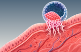
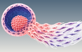
Following preimplantation development, the mammalian blastocyst consists of three principal cell populations: the epiblast, which gives rise to the embryo proper; the hypoblast (primitive endoderm), which contributes primarily to extraembryonic endoderm; and the surrounding trophectoderm, the first lineage specified during embryogenesis. After reaching the uterus, the blastocyst hatches from the zona pellucida and attaches to the receptive endometrial epithelium during the implantation process.
The trophectoderm rapidly differentiates into specialized trophoblast populations that mediate embryo attachment and invasion of the maternal decidua. Two major trophoblast lineages are established:
- Cytotrophoblasts (CTBs): proliferative mononuclear progenitor cells that generate additional trophoblast populations.
- Syncytiotrophoblasts (STBs): multinucleated cells formed through cytotrophoblast fusion that directly contact maternal tissues, facilitate implantation, and initiate maternal–fetal exchange.
As placental development progresses, subsets of cytotrophoblasts further differentiate into extravillous trophoblasts (EVTs), which migrate into the decidua and remodel maternal spiral arteries, thereby establishing adequate uteroplacental blood flow. Together, these trophoblast populations form the structural and functional basis of the developing placenta while regulating communication between maternal and embryonic tissues.
Placenta and Extraembryonic Membranes
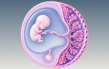
The placenta is a transient but highly specialized extraembryonic organ that performs essential respiratory, nutritional, metabolic, endocrine, and immunological functions throughout gestation. It consists of fetal trophoblast-derived tissues and maternal decidual tissues that together form the maternal–fetal interface. Beyond facilitating nutrient and gas exchange, the placenta synthesizes hormones that support pregnancy, regulates immune tolerance between genetically distinct maternal and fetal tissues, and serves as a selective barrier against pathogens and circulating molecules.
Several extraembryonic membranes develop concurrently with the placenta:
- Amnion: encloses the amniotic cavity and protects the developing embryo within amniotic fluid.
- Yolk sac: supports early nutrient transfer, primitive hematopoiesis, and germ cell development before placental circulation becomes fully established.
- Chorion: contributes the fetal component of the placenta through the formation of chorionic villi.
- Connecting stalk: later develops into the umbilical cord, linking the embryo to the placenta.
Although these structures do not contribute directly to the embryo proper, they are indispensable for normal embryogenesis and fetal development. Their coordinated formation exemplifies the close integration of embryonic and extraembryonic developmental programs during early pregnancy.
The Maternal–Fetal Interface
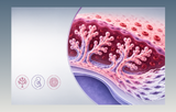
The maternal–fetal interface comprises the dynamic interaction between fetal trophoblast cells and the maternal decidua, immune cells, vasculature, and extracellular matrix. Rather than functioning as a simple physical boundary, this interface coordinates implantation, placental morphogenesis, vascular adaptation, immune regulation, and tissue remodeling throughout gestation.
Successful implantation requires balanced communication between maternal and embryonic tissues that supports trophoblast invasion while maintaining immune tolerance to the semi-allogeneic fetus. Decidual stromal cells, uterine natural killer cells, macrophages, endothelial cells, and trophoblast populations collectively establish an environment that promotes placental development while protecting both maternal and fetal health. Dysregulation of these interactions has been implicated in numerous pregnancy disorders, making the maternal–fetal interface a major focus of reproductive biology and translational research.
Research Tools for Implantation and Placental Biology
Advances in developmental biology research have been driven by increasingly sophisticated experimental platforms that enable investigation of implantation and extraembryonic development in vitro. Researchers rely on validated stem cell culture reagents, defined media, and molecular characterization tools to establish reproducible experimental systems.
Commonly used resources include:
- Extracellular matrices and substrates: recombinant vitronectin, laminin, collagen, and synthetic matrix coatings for pluripotent stem cells and trophoblast cultures.
- Embryonic development antibodies: antibodies against OCT4 (POU5F1), NANOG, SOX2, GATA3, TFAP2C, KRT7, hCG, SSEA-4, TRA-1-60, and TRA-1-81 for lineage identification.
- ESC characterization kits: pluripotency marker panels for immunofluorescence, flow cytometry, quantitative PCR, and gene expression profiling.
- Functional research tools: CRISPR genome-editing reagents, differentiation kits, RNA analysis workflows, single-cell sequencing solutions, live-cell imaging reagents, and validation assays.
Researchers depend on well-characterized, validated reagents to generate reproducible data across studies investigating implantation biology, trophoblast differentiation, placental development, and the maternal–fetal interface. As stem cell technologies, organoid systems, and embryo models continue to evolve, comprehensive collections of specialized culture media, antibodies, characterization assays, and genome engineering tools are enabling increasingly precise investigation of the earliest stages of human development and accelerating discoveries in reproductive biology and regenerative medicine.