



The RayBio® 6-RNA Modification Dot Blot Combination Kit is an in vitro semi-quantitative assay for detecting m1A, m7G, m6A, m5C, ac4C and A-I modifications.
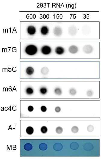
Representative image of 6-RNA modification-specific dot blot assay of RNA samples
6-RNA Modification Dot Blot Combination Kit
Contact Us for Bulk Request
Product Description
Specifications
| Size | 1 Kit |
| Compatible Sample Types | Extracted RNA |
| Quantitative/Semi-Quantitative | Semi-Quantitative |
| Research Area | Epigenetics, RNA/DNA Modification |
| Shipping Type | Blue ice |
| Storage | 4°C |
Related Products
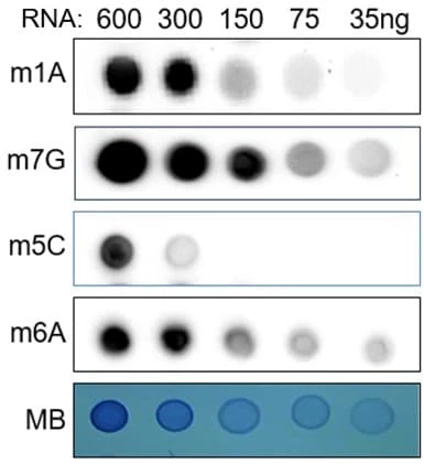
4-RNA Methylation Dot Blot Combination Kit
Introduction
RNA modifications are dynamic and reversible chemical modifications on substrate RNA that are regulated by specific modifying enzymes. They play important roles in the regulation of many biological processes in various diseases, including cancer, neurological disorders, cardiovascular diseases, metabolic diseases, genetic and developmental diseases, as well as immune disorders.
Advanced sequencing technologies have highlighted the significance of RNA modifications in human diseases. Recent studies have mapped out the locations and abundance of key RNA modifications, such as N6-methyladenosine (m6A), 5-methylcytosine (m5C), N1-methyladenosine (m1A), and N7-methylguanosine (m7G), N4-acetylcytidine (ac4C) and A-to-I editing (Adenosine-to-Inosine, A-I) primarily through antibody immunoprecipitation or chemical treatments coupled with next-generation sequencing. It is well known that through the “writing (Methyltransferase) - erasing (Demethyltransferase) -reading (Binding protein)” mechanisms, RNA modifications regulate the stability, translation, and localization of pivotal disease-related mRNAs to manipulate disease development. Further research into RNA modifications, their roles, inhibitors, and activators, is essential for disease diagnosis, treatment, and prognosis.
Here, we introduce a combined m1A, m7G, m6A, m5C, ac4C and A-I dot blot kit, which offers a reliable, simple, and rapid method of quantifying the 6 most common global RNA modification levels in in RNA isolated from cell, tissue, urine, serum, plasma, cell culture medium or other cell samples collected from humans, rats, mice and other species.
Product Features
- Highly sensitive and specific
- Requires minimal sample preparation
- No specialty equipment required
- Suitable for high-throughput screening
- Compatible with various sample types and species
- ≥ 48 samples could be detected by 1 kit
Kit Components
| Component | Size / Description |
|---|---|
| m1A Antibody | 1 vial |
| m7G Antibody | 1 vial |
| m6A Antibody | 1 vial |
| m5C Antibody | 1 vial |
| ac4C Antibody | 1 vial |
| Inosine Antibody | 1 vial |
| HRP-conjugated Secondary Antibody | 1 vial |
| Detection Buffer C | 1 bottle |
| Detection Buffer D | 1 bottle |
| Wash Buffer | 1 bottle |
| Blocking Buffer | 1 bottle |
| MB Staining Buffer | 1 bottle |
| NC Membranes | 4 sheets |
| Plastic Sheet | 8 sheets |
| Incubation Dish | 2 dishes |
Additional Materials Required
- RNase-free tubes
- Heat blocker
- Plastic petri dish
- UV cross-linker
- Chemiluminescent Imaging system
Assay Procedure Summary
- Sample preparation: Depending on the type of sample (RNA, plasma, or serum), different methods of extraction, dilution, and denaturation are required.
- Sample loading and crosslinking: The sample is spotted onto a nitrocellulose membrane, air dried, and crosslinked with UV light twice.
- Methylene blue staining: One line of the spotted sample replicates is cut off and stained with methylene blue buffer to serve as an internal control.
- Blocking and antibody incubation: The membrane is blocked with blocking buffer and then incubated with corresponding antibody and HRP-conjugated secondary antibody.
- Detection and imaging: The membrane is treated with a mixture of detection buffers C and D, which catalyzes a color development reaction. The chemiluminescent signal is captured by an imaging system.
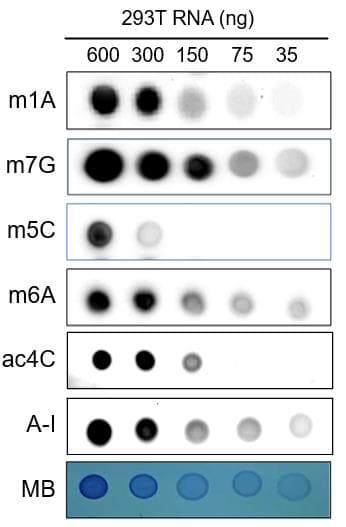
Typical Data
Storage/Stability
The entire kit may be stored at -20°C to -80°C for up to 12 months from the date of shipment. For extended storage, it is recommended to store it at -80°C. Avoid repeated freeze-thaw cycles.