Human CD95/Fas Antibody : LE/AF

Cat# OASB01147
Size : 0.5mg
Brand : Aviva Systems Biology
Contact local distributor :
Human CD95/Fas Antibody : LE/AF (OASB01147)
| Datasheets/Manuals | Printable datasheet for Human CD95/Fas Antibody : LE/AF (OASB01147) |
|---|
| Tested Species Reactivity | Human |
|---|---|
| Predicted Species Reactivity | Human |
| Clonality | Monoclonal |
| Clone | DX2 |
| Isotype | IgG1 |
| Host | Mouse |
| Conjugation | LE: Low Endotoxin, AF: Azide Free |
| Application | FC, IHC-F, IHC-P, ICC, IP |
| Additional Information | Description: CD95, also known as Fas and APO-1, is a 45 kDa type I transmembrane glycoprotein and a member of the tumor necrosis factor receptor superfamily. It is expressed by activated lymphocytes, monocytes, neutrophils, fibroblasts, and cell lines. Fas ligand binding to CD95 induces apoptosis in activated mature lymphocytes thereby playing a role in maintaining peripheral tolerance. Crosslinking of CD95 by the monoclonal antibodies DX2 and DX3 delivers an apoptotic signal to Fas-sensitive cells indicating that these monoclonal antibodies recognize a functional epitope of CD95. |
| Reconstitution and Storage | Store at -20C |
| Immunogen | Human CD95 transfected L cells |
| Concentration | 0.5 mg/mL |
| Specificity | CD95 |
| Characterization | To insure lot to lot consistency, each batch of product is tested by flow cytometry to conform with thecharacteristics of a standard reference. |
| Warning | Reagents contain sodium azide which is very toxic if ingested or inhaled. Avoid contact with skin, eyes, orclothing. Wear eye or face protection when handling. If skin or eye contact occurs, wash with copiousamounts of water. If ingested or inhaled, contact a physician immediately. Sodium azide yields toxichydrazoic acid under acidic conditions. Dilute azide- containing compounds in running water beforediscarding to avoid accumulation of potentially explosive deposits in lead or copper plumbing. |
| Dilution | Flow Cytometry: Purified antibody Fluorescein conjugate Biotin conjugate R-phycoerythrin conjugate Allophycocyanin conjugate <= 1 ug/106 cells 10 uL/106 cells 10 uL/106 cells 10 uL/106 cells 10 uL/106 cells |
| Application Info | Flow cytometry, Immunohistochemistry (frozen sections), Immunoprecipitation, In vitro induction of apoptosis |
| Other Applications Data | Since applications vary, you should determine the optimum workingdilution of the product that is appropriate for your specific need. |
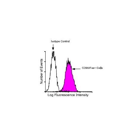
| Other Applications Image 1 Data | Amount Used: 1 Ug/106 cells The Jurkat human lymphoma cell line was stained with mouse anti-human CD95/Fas-FITC, following which large cells were gated and analyzed on a FACScan flow cytometer (BDIS, San Jose, CA). |
| Storage | - The purified (UNLB) antibody is supplied as 0.1 mg of purified immunoglobulin in 1.0 mL of 100 mMborate buffered saline, pH 8.2. No preservatives or amine- containing buffer salts added. Store at 2- 8 C - The fluorescein (FITC) conjugate is supplied as 100 tests in 1.0 mL of PBS/NaN3. Store at 2- 8 C - The biotin (BIOT) conjugate is supplied as 100 tests in 1.0 mL of PBS/NaN3. Store at 2- 8 C - The R- phycoerythrin (R- PE) and allophycocyanin (APC) conjugates are supplied as 100 tests in 1.0mL of PBS/NaN3 and a stabilizing agent. Store at 2- 8 C. Do not freeze! - The low endotoxin/azide- free antibody is supplied as 0.5 mg purified immunoglobulin in 1.0 mL of PBS;aliquot and freeze upon receipt - Protect conjugated forms from light. Reagents are stable for the period shown on the label if stored asdirected. |
| Reference | 1. Schlossman, S., L. Bloumsell, W. Gilks, J.M. Harlan, C. Kishimoto, J. Ritz, S. Shaw, R. Silverstein, T. Springer, T.F. Tedder, and R.F. Todd,eds. 1995. Leukocyte Typing V: White Cell Differentiation Antigens, Oxford University Press, Oxford 2. Kishimoto, T., A.E.G. von dern Borne, S.M. Goyert, D.Y. Mason, M. Miyasaka, L. Moretta, K. Okumura, S. Shaw, T.A. Springer, K Sugamura, and H. Zola, eds. 1998. Leukocyte Typing VI: White Cell Differentiation Antigens, Academic Press, New York 3. Barclay, A.N., M.H. Brown, S.K.A. Law, A.J. McKnight, M.G. Tomlinson, and P.A. van der Merwe, eds. 1997. The Leukocyte AntigensFacts Book, 2nd Edition, CD95 Section, Academic Press, New York, p. 363 4. Nagata, S., and P. Golstein. 1995. Science 267:3378 5. van Parijs, L., and A.K. Abbas. 1996. Curr. Opin. Immunol. 8:355. |
|---|---|
| Gene Symbol | FAS |
| Gene Full Name | Tumor necrosis factor receptor superfamily member 6 |
| Alias Symbols | APT1, CD95, FAS1, APO-1, FASTM, ALPS1A, TNFRSF6 |
| NCBI Gene Id | 355; 574332; 103216224; 105587157 |
| Protein Name | tumor necrosis factor receptor superfamily member 6 |
| Description of Target | The protein encoded by this gene is a member of the TNF-receptor superfamily. This receptor contains a death domain. It has been shown to play a central role in the physiological regulation of programmed cell death, and has been implicated in the pathogenesis of various malignancies and diseases of the immune system. The interaction of this receptor with its ligand allows the formation of a death-inducing signaling complex that includes Fas-associated death domain protein (FADD), caspase 8, and caspase 10. The autoproteolytic processing of the caspases in the complex triggers a downstream caspase cascade, and leads to apoptosis. This receptor has been also shown to activate NF-kappaB, MAPK3/ERK1, and MAPK8/JNK, and is found to be involved in transducing the proliferating signals in normal diploid fibroblast and T cells. Several alternatively spliced transcript variants have been described, some of which are candidates for nonsense-mediated mRNA decay (NMD). The isoforms lacking the transmembrane domain may negatively regulate the apoptosis mediated by the full length isoform. |
| Uniprot ID | P25445, Q9BDP2, Q9BDN4 |
| Protein Accession # | NP_000034.1 |
| Nucleotide Accession # | NM_000043.5 |