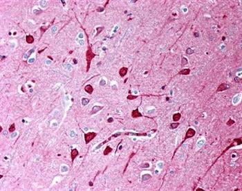
Immunohistochemistry staining of human brain showing labelling of neurons with T8250-16E.
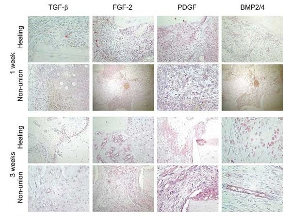
Growth factor expression in healing and non-union groups using T8250-16E. Immunohistochemical analysis of TGF-beta, FGF-b, PDGF and BMP2/4 in healing and non-union groups at 1 and 3 weeks post-osteotomy. Images taken at ×20 or ×40 magnification and positive growth factor staining is seen as a red signal.
For Research Use Only. Not for use in diagnostic procedures.Catalog #: MBS6248369
MBS6248369 is an antibody targeting TGFB1. Also known as Transforming Growth Factor beta 1 (TGFb1, TGF-beta-1, TGFB1, TGFB) (AP). Reactive in Bovine, Human, Mouse, Rabbit, Rat, Sheep. Host: Mouse. More details
Supplied as a liquid in PBS, pH 7.2. Labeled with Alkaline Phosphatase.
Purity/Purification
Purified by Affinity Chromatography
Specificity
Recognizes human platelet-derived and recombinant Transforming Growth Factor beta 1 in ELISA. Demonstrates neutralising activity against TGF-beta1 in cell proliferation assays. Demonstrated to react with dimeric (25kD) or monomeric (12.5kD) molecules of natural TGFb1 under non-reducing and reducing conditions respectively. Species Crossreactivity: rat, sheep, mustelid, rabbit, mouse, bovine and mink
Human Transforming Growth Factor beta 1 from platelets.
Conjugate
AP
Product Categories/Family for anti-TGFB1 antibody
Antibodies; Growth Factors; Cytokines
Applications Tested/Suitable for anti-TGFB1 antibody
ELISA (EIA), Immunohistochemistry (IHC), Western Blot (WB)
Immunohistochemistry (IHC) of anti-TGFB1 antibody
Immunohistochemistry staining of human brain showing labelling of neurons with T8250-16E.
Immunohistochemistry (IHC) of anti-TGFB1 antibody
Growth factor expression in healing and non-union groups using T8250-16E. Immunohistochemical analysis of TGF-beta, FGF-b, PDGF and BMP2/4 in healing and non-union groups at 1 and 3 weeks post-osteotomy. Images taken at ×20 or ×40 magnification and positive growth factor staining is seen as a red signal.
Related Product Information for anti-TGFB1 antibody
MBS6248369 is an antibody targeting TGFB1. Also known as Transforming Growth Factor beta 1 (TGFb1, TGF-beta-1, TGFB1, TGFB) (AP). Reactive in Bovine, Human, Mouse, Rabbit, Rat, Sheep. Host: Mouse.
Product References and Citations for anti-TGFB1 antibody
1. Lehr, E.J.et al. (2010) Decellularization reduces immunogenicity of sheep pulmonary artery vascular patches. J Thorac Cardiovasc Surg. Jul 14. 2. Westermann, D. et al. (2011) Circ Heart Fail. 4: 44-52. 3. Brown, H. et al. (1999) J Infect Dis. 180: 1742-6. 4. Helske, S. et al. (2006) Eur Heart J. 27: 1495-504. 5. Hsiao, Y.W. et al. (2004) J Immunol. 172: 1508-14. 6. Kingston, P.A. et al. (2003) Circulation. 108: 2819-25. 7. Lavaud, S. et al. (2001) Am J Physiol Renal Physiol. 280: F683-94. 8. Sheu, B.C. et al. (2001) J Immunol. 167: 2972-8. 9. Hopkinson, A. et al. (2006) Invest Ophthalmol Vis Sci. 47: 4316-22. 10. Sumpter, T.L. et al. (2007) J Leukoc Biol. 83: 708-17. 11. Braun, N. et al. (2011) Perit Dial Int. 31: 291- 300. 12. Shi, W.K. et al. (1993) [Biological characterization of a monoclonal antibody TB 21 against human transforming growth factor-beta 1]. Shi Yan Sheng Wu Xue Bao. 26: 141-50.
Other Notes
Small volumes of anti-TGFB1 antibody vial(s) may occasionally become entrapped in the seal of the product vial during shipment and storage. If necessary, briefly centrifuge the vial on a tabletop centrifuge to dislodge any liquid in the container`s cap. Certain products may require to ship with dry ice and additional dry ice fee may apply.
Additional Details
NCBI/Uniprot data below describe general gene information for TGFB1. It may not necessarily be applicable to this product.
This gene encodes a member of the transforming growth factor beta (TGFB) family of cytokines, which are multifunctional peptides that regulate proliferation, differentiation, adhesion, migration, and other functions in many cell types. Many cells have TGFB receptors, and the protein positively and negatively regulates many other growth factors. The secreted protein is cleaved into a latency-associated peptide (LAP) and a mature TGFB1 peptide, and is found in either a latent form composed of a TGFB1 homodimer, a LAP homodimer, and a latent TGFB1-binding protein, or in an active form composed of a TGFB1 homodimer. The mature peptide may also form heterodimers with other TGFB family members. This gene is frequently upregulated in tumor cells, and mutations in this gene result in Camurati-Engelmann disease.[provided by RefSeq, Oct 2009]
TGFB1: Multifunctional protein that controls proliferation, differentiation and other functions in many cell types. Many cells synthesize TGFB1 and have specific receptors for it. It positively and negatively regulates many other growth factors. It plays an important role in bone remodeling as it is a potent stimulator of osteoblastic bone formation, causing chemotaxis, proliferation and differentiation in committed osteoblasts. Homodimer; disulfide-linked, or heterodimer with TGFB2. Secreted and stored as a biologically inactive form in the extracellular matrix in a 290 kDa complex (large latent TGF-beta1 complex) containing the TGFB1 homodimer, the latency-associated peptide (LAP), and the latent TGFB1 binding protein-1 (LTBP1). The complex without LTBP1 is known as the'small latent TGF-beta1 complex'. Dissociation of the TGFB1 from LAP is required for growth factor activation and biological activity. Release of the large latent TGF-beta1 complex from the extracellular matrix is carried out by the matrix metalloproteinase MMP3. May interact with THSD4; this interaction may lead to sequestration by FBN1 microfibril assembly and attenuation of TGFB signaling. Interacts with the serine proteases, HTRA1 and HTRA3: the interaction with either inhibits TGFB1-mediated signaling. The HTRA protease activity is required for this inhibition. Interacts with CD109, DPT and ASPN. Activated in vitro at pH below 3.5 and over 12.5. Highly expressed in bone. Abundantly expressed in articular cartilage and chondrocytes and is increased in osteoarthritis (OA). Co-localizes with ASPN in chondrocytes within OA lesions of articular cartilage. Belongs to the TGF-beta family.
Protein type: Motility/polarity/chemotaxis; Secreted; Secreted, signal peptide
Molecular Function: protein binding; protein homodimerization activity; enzyme binding; growth factor activity; protein heterodimerization activity; punt binding; cytokine activity; protein N-terminus binding; glycoprotein binding; antigen binding
Biological Process: extracellular matrix organization and biogenesis; positive regulation of apoptosis; positive regulation of transcription, DNA-dependent; SMAD protein nuclear translocation; female pregnancy; positive regulation of protein amino acid dephosphorylation; activation of NF-kappaB transcription factor; regulation of protein import into nucleus; positive regulation of MAP kinase activity; connective tissue replacement during inflammatory response; regulation of transforming growth factor beta receptor signaling pathway; negative regulation of ossification; cell cycle arrest; inner ear development; positive regulation of isotype switching to IgA isotypes; regulatory T cell differentiation; response to drug; positive regulation of interleukin-17 production; positive regulation of chemotaxis; positive regulation of smooth muscle cell differentiation; active induction of host immune response by virus; positive regulation of blood vessel endothelial cell migration; regulation of sodium ion transport; negative regulation of blood vessel endothelial cell migration; negative regulation of fat cell differentiation; lymph node development; positive regulation of protein secretion; positive regulation of transcription from RNA polymerase II promoter; response to progesterone stimulus; endoderm development; myelination; positive regulation of odontogenesis; negative regulation of phagocytosis; evasion of host defenses by virus; positive regulation of cellular protein metabolic process; myeloid dendritic cell differentiation; negative regulation of transcription from RNA polymerase II promoter; phosphate metabolic process; negative regulation of cell proliferation; negative regulation of T cell proliferation; ureteric bud development; regulation of DNA binding; negative regulation of release of sequestered calcium ion into cytosol; salivary gland morphogenesis; positive regulation of cell proliferation; protein kinase B signaling cascade; protein export from nucleus; inflammatory response; positive regulation of exit from mitosis; aging; epidermal growth factor receptor signaling pathway; mitotic cell cycle checkpoint; common-partner SMAD protein phosphorylation; positive regulation of phosphoinositide 3-kinase activity; positive regulation of bone mineralization; positive regulation of peptidyl-serine phosphorylation; SMAD protein complex assembly; positive regulation of protein kinase B signaling cascade; positive regulation of protein complex assembly; positive regulation of protein import into nucleus; response to hypoxia; epithelial to mesenchymal transition; negative regulation of cell growth; negative regulation of cell-cell adhesion; negative regulation of transforming growth factor beta receptor signaling pathway; negative regulation of skeletal muscle development; mononuclear cell proliferation; regulation of cell migration; protein amino acid phosphorylation; hyaluronan catabolic process; regulation of apoptosis; response to vitamin D; negative regulation of neuroblast proliferation; receptor catabolic process; positive regulation of superoxide release; transforming growth factor beta receptor signaling pathway; germ cell migration; response to glucose stimulus; chondrocyte differentiation; defense response to fungus, incompatible interaction; T cell homeostasis; negative regulation of mitotic cell cycle; cell growth; tolerance induction to self antigen; regulation of striated muscle development; platelet activation; organ regeneration; negative regulation of DNA replication; virus-host interaction; hemopoietic progenitor cell differentiation; negative regulation of transcription, DNA-dependent; positive regulation of epithelial cell proliferation; positive regulation of collagen biosynthetic process; viral infectious cycle; response to estradiol stimulus; negative regulation of cell cycle; positive regulation of histone deacetylation; response to radiation; platelet degranulation; negative regulation of protein amino acid phosphorylation; lipopolysaccharide-mediated signaling pathway; response to wounding; adaptive immune response based on somatic recombination of immune receptors built from immunoglobulin superfamily domains; negative regulation of epithelial cell proliferation; intercellular junction assembly and maintenance; regulation of binding; MAPKKK cascade; cellular calcium ion homeostasis; gut development; protein import into nucleus, translocation; ATP biosynthetic process; positive regulation of histone acetylation; positive regulation of protein amino acid phosphorylation; negative regulation of myoblast differentiation; blood coagulation; positive regulation of cell migration
All of MyBioSource's Products are for scientific laboratory research purposes and are not for diagnostic, therapeutics, prophylactic or in vivo use. Through your purchase, you expressly represent and warrant to MyBioSource that you will properly test and use any Products purchased from MyBioSource in accordance with industry standards. MyBioSource and its authorized distributors reserve the right to refuse to process any order where we reasonably believe that the intended use will fall outside of our acceptable guidelines.
Disclaimer
While every efforts were made to ensure the accuracy of the information provided in this datasheet, MyBioSource will not be liable for any omissions or errors contained herein. MyBioSource reserves the right to make changes to this datasheet at any time without prior notice.
It is the responsibility of the customer to report product performance issues to MyBioSource within 30 days of receipt of the product. Please visit our Terms & Conditions page for more information.
Product Note
Select online data sheet information is drawn from bioinformatics databases, occasionally resulting in ambiguous or non-relevant product information. It is the responsibility of the customer to review, verify, and evaluate the information to make sure it matches their requirements before purchasing the kit. Our ELISA Kit assays are dynamic research tools and sometimes they may be updated and improved. If the format of this assay is important to you then please request the current manual or contact our technical support team with a presales inquiry before placing an order. We will confirm the current details of the assay. We cannot guarantee the sample manual posted online is the most current manual, it is intended to serve as an example only. Please refer to the instructions For Use provided with the assay kit for precise details.