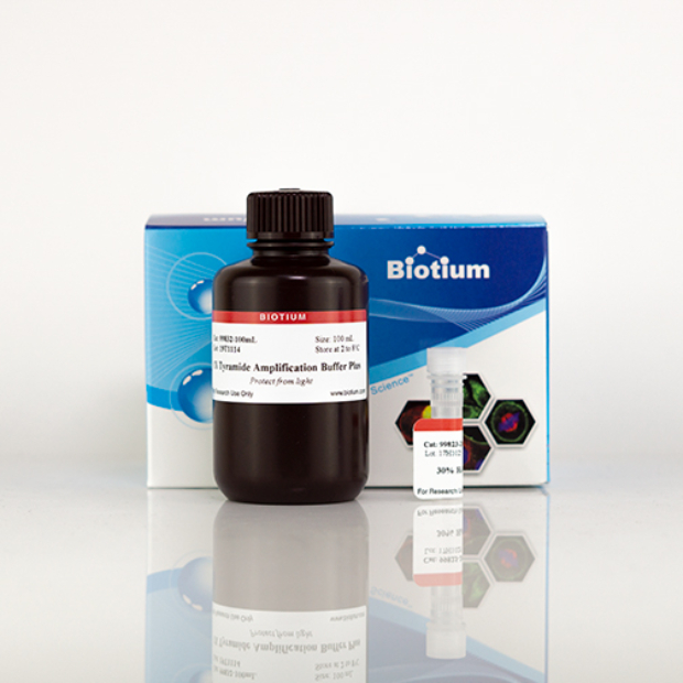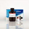
Tyramide Amplification Buffer Plus, 250 reactions
Cat# 22029-T
Size : 1kit
Brand : Biotium
Tyramide Amplification Buffer Plus
An optimized buffer with enhanced sensitivity for tyramide signal amplification of immunostaining in cells or tissue sections.

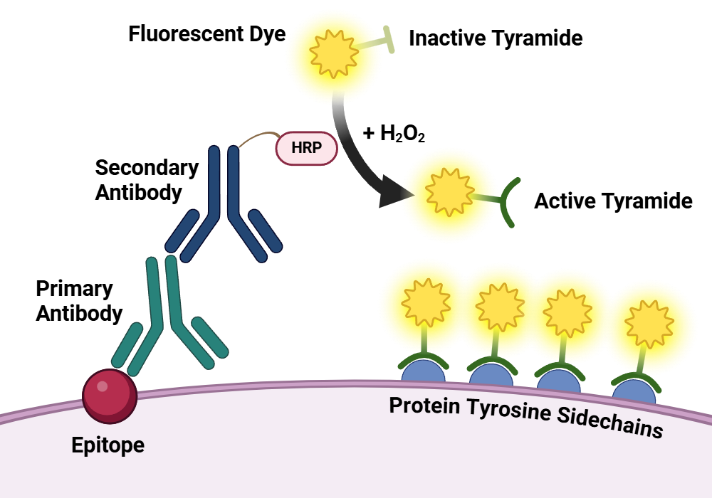
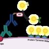
Illustration of the tyramide signal amplification system. A cell or tissue sample is labeled with primary and secondary antibody using conventional methods. The horseradish peroxidase, conjugated to the secondary antibody, catalyzes the conversion of labeled tyramide into a reactive radical. The tyramide radical then covalently binds to nearby tyrosine residues, providing high-density labeling. Created in https://BioRender.com.
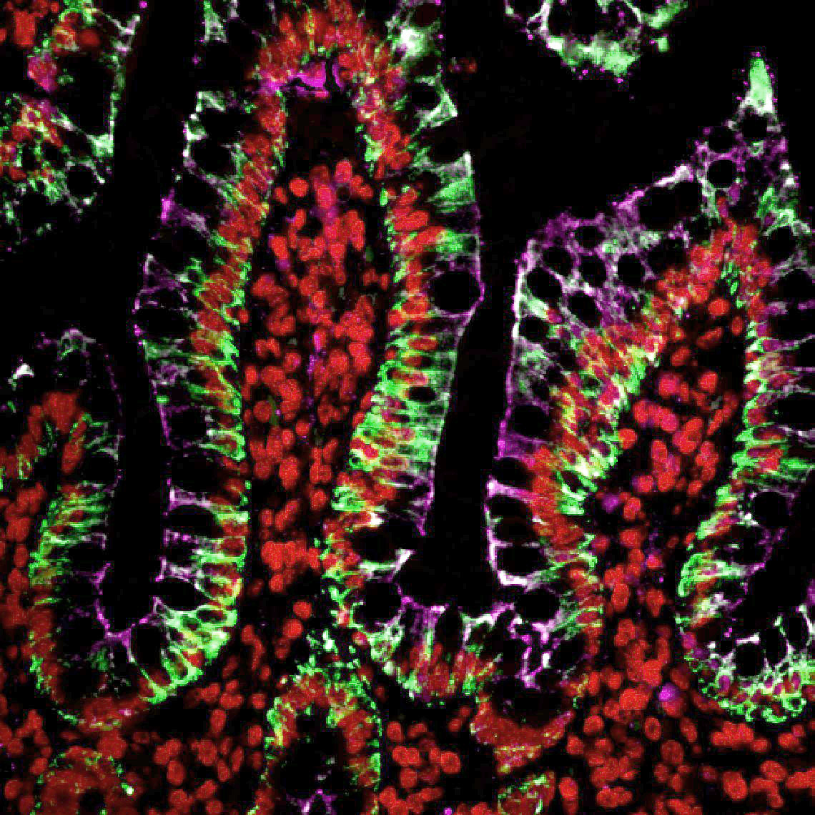
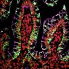
Multiplex tyramide labeling of FFPE tissues. CF®488A-tyramide labeling pan-CK (green); Cy®3-tyramide labeling histone H1 (red); CF®640R-tyramide labeling ZO1 (magenta). Primary antibodies were from mouse, and secondary antibody was HRP-conjugated goat anti-mouse. Each labeling was performed sequentially, with antibody removal by microwave treatment.
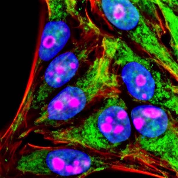
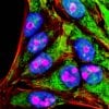
Sequential labeling of formaldehyde-fixed HeLa cells with two Tyramide Amplification Kits. Mitochondria (green) were imaged with rabbit anti-COXIV primary antibody and Tyramide Amplification Kit with HRP goat anti-rabbit IgG and CF®488A-tyramide, followed by peroxidase quenching. Nucleoli (magenta) were visualized with mouse anti-cyclin B1 primary antibody and Tyramide Amplification Kit with HRP Goat anti-mouse IgG and CF®568-tyramide. Actin (red) was detected with CF®640R-phalloidin, and cell nuclei (blue) were stained with DAPI.
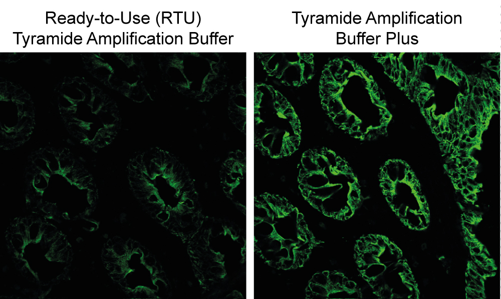
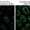
PanCK staining in human colon cryosections detected using CF®488A tyramide with Ready-to-Use Tyramide Amplification Buffer (left), or Tyramide Amplification Buffer Plus (right). Amplification Buffer Plus gives brighter signal compared to original Ready-to-Use Tyramide Amplification Buffer.
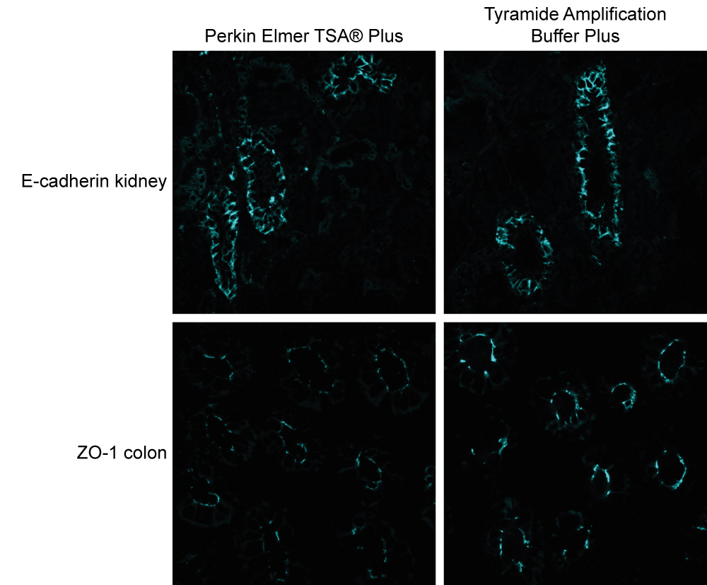
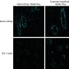
Staining of E-cadherin in human kidney (top) and ZO1 in human colon (bottom) cryosections using CF®405S tyramide. Cryosections were also treated with Perkin Elmer's TSA® Plus Reagent (left) and Biotium's Tyramide Amplification Buffer Plus (right). Amplification Buffer Plus gives brighter signal compared to Perkin Elmer's TSA® Plus Reagent.
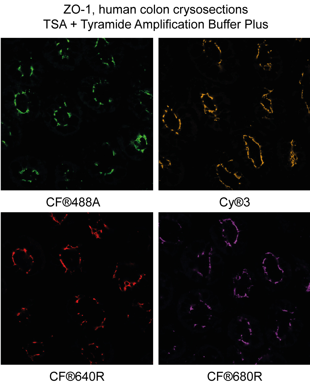
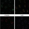
ZO-1 staining in human colon cryosections detected using CF®488A, Cyanine 555 (Cy®3), CF®640R, and CF®680R tyramide with Biotium's Tyramide Amplification Buffer Plus.
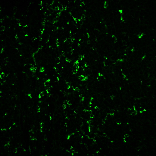
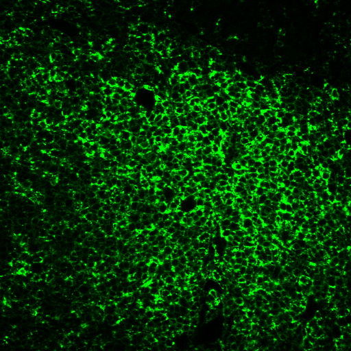
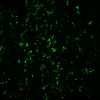
Human tonsil FFPE section stained with anti-CD68 (C68/684+KP1) and goat anti-mouse HRP, detected using CF®488A tyramide in Tyramide Amplification Buffer Plus.
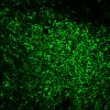
Human tonsil FFPE section stained with anti-CD45RO (UCHL-1) and goat anti-mouse HRP, detected using CF®488A tyramide in Tyramide Amplification Buffer Plus.
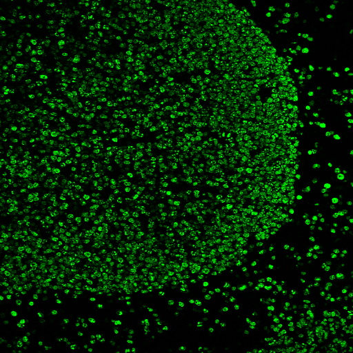
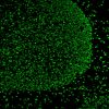
Human tonsil FFPE sections heated in AntiFix™ antigen retrieval buffer, then stained with anti-Ki67 MKI67/2466, goat anti-mouse HRP, and CF®488A tyramide using Tyramide Amplification Buffer Plus.Product Description
Tyramide Amplification Buffer Plus is an optimized buffer with enhanced sensitivity for tyramide signal amplification of immunostaining in cells or tissue sections. It provides significantly improved brightness, specificity, and sensitivity compared to our original Ready-to-Use Tyramide Amplification Buffer (22027).
Features
- For cell and tissue staining with tyramide signal amplification
- Significantly improved brightness, specificity, and sensitivity
- Compatible with CF® Dye & other tyramides
- 250 or 1000 reaction sizes*
* With 100 uL reaction volume; number of reactions may vary depending on sample/specimen size.
Components
- 1X Tyramide Amplification Buffer Plus
- 30% Hydrogen Peroxide
Tyramide Amplification Buffer Plus was developed as a new and improved version of our original Ready-to-Use Tyramide Amplification Buffer (22027). It is also a component in all of our Tyramide Amplification Kits. To learn more about tyramide signal amplification, including our Tyramide Amplification Kits with a selection of bright and photostable CF® dyes, see the Tyramide Amplification Technology Page.


Powered by Bioz