
Zika virus Envelope protein antibody

This image was provided courtesy of cooperative research laboratories.
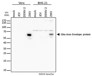
Mock and infected Vero and BHK-21 whole cell extracts (20 μg) were separated by gradient gel, and the membrane was blotted with Zika virus Envelope protein antibody (GTX133314) diluted at 1:4000.
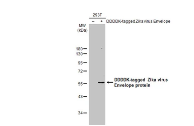
Non-transfected (–) and transfected (+) 293T whole cell extracts (60 μg) were separated by 10% SDS-PAGE, and the membrane was blotted with Zika virus Envelope protein antibody (GTX133314) diluted at 1:5000. The HRP-conjugated anti-rabbit IgG antibody (GTX213110-01) was used to detect the primary antibody.
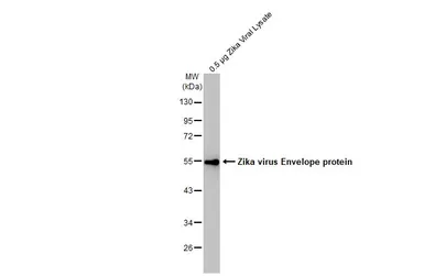
Zika viral lysate (0.5 μg) was separated by 10% SDS-PAGE, and the membrane was blotted with Zika virus Envelope protein antibody (GTX133314) diluted at 1:2000. The HRP-conjugated anti-rabbit IgG antibody (GTX213110-01) was used to detect the primary antibody.
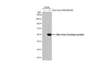
Non-infected (–) and infected (+) C6/36 whole cell extracts (30 μg) were separated by 10% SDS-PAGE, and the membrane was blotted with Zika virus Envelope protein antibody (GTX133314) diluted at 1:1000. The HRP-conjugated anti-rabbit IgG antibody (GTX213110-01) was used to detect the primary antibody.
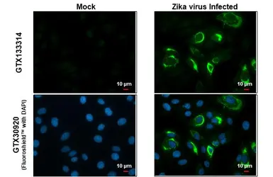
Immunofluorescent analysis of Zika virus infected cells using Zika virus NS5 protein antibody (GTX133314).Sample: Mock and zika virus-infected cells.Green: Zika virus Envelope protein antibody (GTX133314) diluted at 1:100.
This image was provided courtesy of cooperative research laboratories.
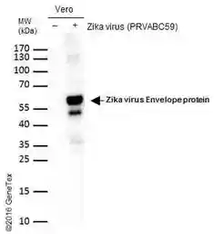
Non-infected (–) and infected (+) vero cells (15 μg) were separated by gradient gel, and the membrane was blotted with Zika virus Envelope protein antibody (GTX133314) diluted at 1:2000. The HRP-conjugated anti-rabbit IgG antibody (GTX213110-01) was used to detect the primary antibody.

This image was provided courtesy of cooperative research laboratories.
Immunofluorescent analysis of non-infected and infected vero or BHK-21 cells using Zika virus Envelope protein antibody (GTX133314).
Green: Zika virus Envelope protein antibody (GTX133314) diluted at 1:4000.
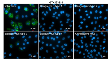
Immunofluorescent analysis of arboviruses infected cells using Zika virus Envelope protein antibody (GTX133314).
Samples: EUROIMMUN Arboviral Fever Mosaic 2 slide (FR 2668-1010-1).
Green: Zika virus Envelope protein antibody (GTX133314) diluted at 1:500.
Blue: Hoechst 33342 staining.
Scale bar = 10 μm.

This image was provided courtesy of cooperative research laboratories.
Immunofluorescent analysis of Zika Virus-PRVABC59 infected (A) and non-infected (B) vero cells using Zika virus Envelope protein antibody (GTX133314).
Green: Zika virus Envelope protein antibody (GTX133314) diluted at 1:4000.
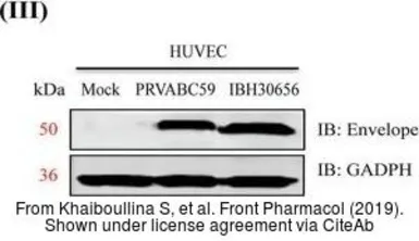
The data was published in the journal Front Pharmacol in 2019. PMID: 31249527
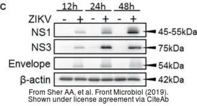
The data was published in the journal Front Microbiol in 2019. PMID: 30984137
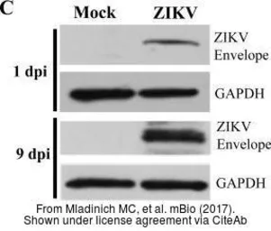
The data was published in the journal mBio in 2017. PMID: 28698279
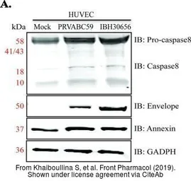
The data was published in the journal Front Pharmacol in 2019. PMID: 31249527
The data was published in the journal Emerg Microbes Infect in 2019. PMID: 30866755
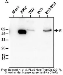
The data was published in the journal PLoS Negl Trop Dis in 2017.PMID: 28481898
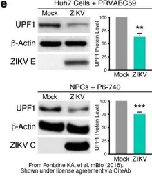
The data was published in the journal MBio in 2018.PMID: 30401782
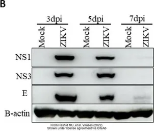
The data was published in the 2022 in Viruses. PMID: 35215967