Key Features of CE/IVD Dermatopathology Primary Antibodies
- CE/IVD-compliant reagents intended for in vitro diagnostic applications in clinical pathology laboratories.
- Validated performance on formalin-fixed, paraffin-embedded (FFPE) tissue specimens.
- Designed to provide high analytical performance when used according to the manufacturer's validated protocols and instructions for use (IFUs).
- Optimized staining protocols compatible with heat-induced epitope retrieval (HIER) methods.
- Availability in ready-to-use and concentrated formats to support laboratory workflow flexibility.
- Reproducible staining performance with optimized signal-to-background characteristics under validated assay conditions.
- Compatibility with automated IHC staining platforms and standardized laboratory workflows, depending on assay validation and platform specifications.
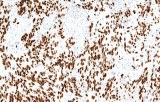
- Support for the interpretation of nuclear, cytoplasmic, membranous, and other diagnostically relevant subcellular staining patterns.
- Lot-to-lot consistency supported by rigorous quality-control procedures and validation testing.
- Comprehensive technical documentation, including instructions for use (IFUs), recommended controls, and interpretation guidance.