Adhesive and coated slides for immunohistochemistry (IHC) and in situ hybridization (ISH) are essential tools for scientists and researchers engaged in histological and molecular pathology. These slides provide the critical surface to which tissue specimens are firmly adhered, enabling precise analysis of tissue morphology, protein markers, and nucleic acid localization. They enhance specimen retention, prevent tissue loss during staining protocols, and improve signal quality, making them indispensable in cancer research, biomarker discovery, and diagnostic applications.
Importance of Adhesive and Coated Slides in IHC and ISH
-
Adhesive slides are treated with chemical coatings that increase tissue adhesion by creating a hydrophilic or charged surface compatible with formalin-fixed, paraffin-embedded (FFPE) or frozen specimens. Common coatings include poly-L-lysine, silane, and proprietary formulations optimized for different tissue types and staining procedures.
-
Coated slides help maintain tissue integrity during multiple washes and antibody or probe incubations, which are routine in IHC and ISH protocols.
-
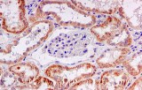
These slides support high sensitivity and reproducibility in detecting protein antigens or nucleic acids, essential for accurate pathological interpretation and research conclusions.
-
Specialized coated slides such as polyethylene naphthalate (PEN)-membrane slides can facilitate laser capture microdissection for downstream genomics or proteomics analysis.
Features and Benefits
-
Enhanced adhesion reduces tissue detachment and loss, ensuring consistent and reliable data.
-
Compatibility with multiple staining procedures, including multiplex IHC and ISH workflows.
-
High optical clarity and low autofluorescence for superior imaging quality.
-
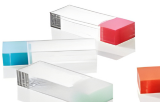
Available in various formats: standard glass slides, charged slides, membrane-coated slides, and slides designed for automation compatibility.
-
Some advanced coatings are optimized for heat-induced epitope retrieval (HIER), preserving antigenicity while securing tissue.
Application Context and Scientific Relevance
-
Widely used in clinical diagnostics, cancer research, and molecular pathology to detect biomarkers for targeted therapies.
-
Integral for studies involving tumor microenvironment profiling, immune cell phenotyping, and gene expression analysis.
-
Suitable for cutting-edge multiplex immunohistochemistry and immunofluorescence platforms enabling simultaneous detection of multiple targets in a single tissue section.
-
Support ongoing advances in digital pathology and whole-slide imaging through consistent sample preparation quality.